Anatomy of the Dog Images
Brain Chapter 18
http://vanat.cvm.umn.edu/brain18/
Return to Menu Page
This Page Displays Finished Images & Print PDFs
(Last updated: 3/22/11)
Alvin J. Beitz, PhD and Thomas F. Fletcher, DVM, PhD
Department of Veterinary & Biomedical Sciences
College of Veterinary Medicine
University of Minnesota
235c AS/VM Bldg.
St. Paul MN 55108
*************
Phone (612) 624-9765
Fax: (612) 625-0204
e-mail: fletc003@umn.edu
Note:
This web site is intended to update book editors regarding image development progress for Anatomy of the Dog Chapter 18 Brain and to obtain editor comments. Below is an unordered list of the images that have been created so far. Click a listed image to view an actual-size version of the image and its caption in a pop-up web page. Also, a link to download a PDF print version of each image is provided, in case it is needed. (Final versions of the images will be submitted to Saunders as TIFF or EPS documents.)
Assumptions & parameters we are using for chapter images:
- Book page width will be 7 inches; column width will be 3 & 3/8 inches.
- Cartoon images have been created as vector drawings using Adobe Illustrator and they are high resolution.
- Adobe Photoshop pixel-map image files will be available for submission as TIFF final products. The Photoshop files are CYMK 300 ppi with additional high-resolution layers for label lines and each text label (typically Helvetica).
- The PDF image files that can be downloaded from this site are intended for printing to evaluate image size and quality. Quality should be comparable to the Photoshop files from which the PDFs were generated.
- The screen images shown on this web site have been converted to RGB, and optimized for the web. The screen images are intended for previewing captions, images, labels, placements, etc.
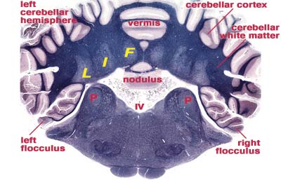
Cerebellar Nuclei
|
Click the image to view an enlarged version and caption in a separate window. To download a PDF print version of the image, click here. The PDF file is approximately 19 MB in size. (Updated 6/30/10) Fig. 46 |
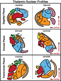
Thalamic Nuclei
|
Click the image to view an enlarged version and caption in a separate window. To download a PDF print version of the image, click here. The PDF file is approximately 300 KB in size. (Updated 11/24/10[caption]) Fig. 23 |
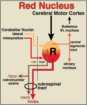
Red Nucleus
|
Click the image to view an enlarged version and caption in a separate window. To download a PDF print version of this column-width image, click here. The PDF file is approximately 0.7 MB in size. (Updated 3/22/11) Fig. 20 |
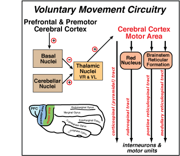
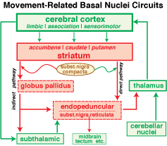
Vountary Movement Circuit
|
Click the image to view an enlarged version and caption in a separate window. To download a PDF print version of this column-width image, click here. The PDF file is approximately 0.8 MB in size. (Updated 3/16/11) Fig. 43 |
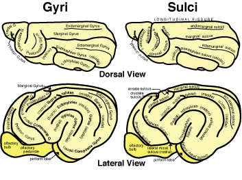
Sulci & Gyri
|
Click the image to view an enlarged version and caption in a separate window. To download a PDF print version of the image, click here. The PDF file is approximately 400 KB in size. (Updated 3/11/11) Fig. 30 |
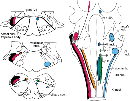
Cranial Nerve Nuclei
|
Click the image to view an enlarged version and caption in a separate window. To download a PDF print version of the image, click here. The PDF file is approximately 250 KB in size. (Updated 6/30/10) Fig. 5 |
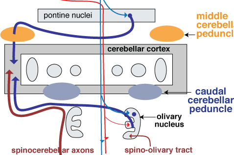
Cerebellar Connections
|
Click the image to view an enlarged version and caption in a separate window. To download a PDF print version of the image, click here. The PDF file is approximately 250 KB in size. (Updated 7/12/10) Fig. 49 |
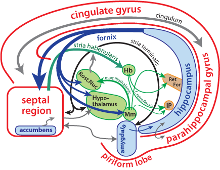
Limbic System Components
|
Click the image to view an enlarged version and caption in a separate window. To download a PDF print version of the image, click here. The PDF file is approximately 280 KB in size. (Updated 8/16/10) Fig. 39 |
Brainstem Reticular Nuclei
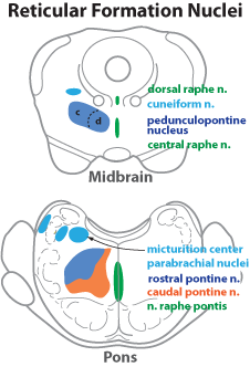
|
Click the image to view an enlarged version and caption in a separate window. To download a PDF print version of this column-width image, click here. The PDF file is approximately 250 KB in size. (Updated 8/25/10) Fig. 6 |
Brainstem Visceral Nuclei
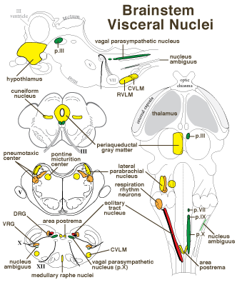
|
Click the image to view an enlarged version and caption in a separate window. To download a PDF print version of the image, click here. The PDF file is approximately 240 KB in size. (Updated 8/26/10) Fig. 7 |
Brainstem Neuromodulatory Nuclei
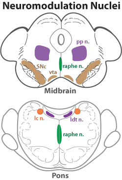
|
Click the image to view an enlarged version and caption in a separate window. To download a PDF print version of this column-width image, click here. The PDF file is approximately 250 KB in size. (Updated 8/26/10) Fig. 15 |
Basal Nuclei
|
|
Click the image to view an enlarged version and caption in a separate window. To download a PDF print version of the image, click here. The PDF file is approximately 1.3 in size. (Updated 4/11/11) Fig. 44 |
Basal Nuclei
|
Click the image to view an enlarged version and caption in a separate window. To download a PDF print version of the image, click here. The PDF file is approximately 0.7 MB in size. (Updated 11/18/10) Fig. 45 |
Hypothalamic Nuclei

|
Click the image to view an enlarged version and caption in a separate window. To download a PDF print version of the image, click here. The PDF file is approximately 33 MB in size. (Updated 12/13/10) Fig. 25 |
Cerebellar Lobes

|
|
Embryonic Brain Divisions
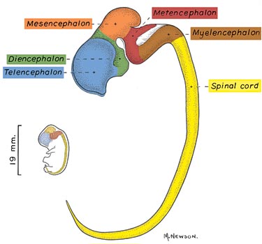
|
|
Hippocampal Formation
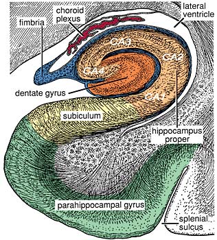
|
|
Neocortical Layers
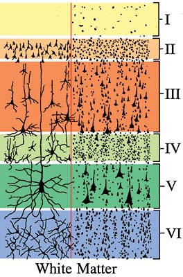
|
|
Cortical Regions
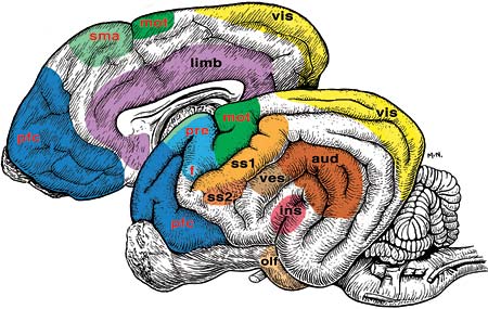
|
|
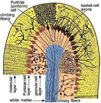
Cerebellar Folium.
 |
|
Go Top