Neocortical Layers
CLOSE
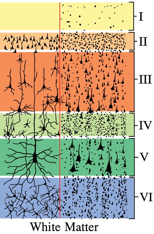
Figure 18—41. Schematic illustration of the six horizontal layers characteristic of the cerebral neocortex. Layers vary in thickness among different regions of cortex. The left side show individual cell profiles as would be seen in a Golgi stain. The right side shows populations of cell body profiles as would be seen in a Nissl stain. I = molecular layer; II = outer granular layer; III = outer pyramidal layer; IV = inner granular layer; V = inner pyramidal layer; and VI = multiform layer. (Modified from Ranson, S.W. and Clark S.L. 1959 Anatomy of the Nervous System, 10th ed. Philadelphia, W. B. Saunders Co.)
Go Top