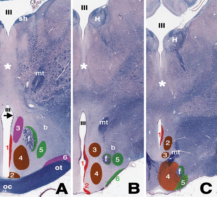
Figure 18—25. Approximate locations of hypothalamic nuclei are illustrated schematically at three transverse levels (purple = axons to neurohypophysis; red = periventricular zone; brown/orange = medial zone; green = lateral zone). A. Rostral level, through optic chiasm: 1 = periventricular nucleus; 2 = suprachiasmatic nucleus; 3 = paraventricular nucleus; 4 = rostral hypothalamic nucleus; 5 = lateral hypothalamic nucleus; 6 = supraoptic nucleus; f = perifornical nucleus (green) around the fornix; (preoptic nuclei are rostral to this level). The arrow indicates the hypothalamic sulcus, ventral to which the hypothalamic vascular organ is evident. B. Intermediate level, through the tuber cinereum: 1 = periventricular nucleus; 2 = infundibular (arcuate) nucleus; 3 = dorsomedial nucleus; 4 = ventromedial nucleus; 5 = lateral hypothalamic nucleus; 6 = tuberal nuclei; f = perifornical nucleus (green) around the fornix. C. Caudal level, through the mamillary body: 1 = periventricular nucleus; 2 = caudal hypothalamic nucleus; 3 = supramamillary nucleus; 4 = medial mamillary nucleus; 5 = lateral mamillary nucleus; f = perifornical nucleus (green) around the fornix. For all levels: * = interthalamic adhesion; III = third ventricle; b = medial forebrain bundle; f = column of fornix; H = habenula; mt = mamillothalamic tract; oc = optic chiasm; ot = optic tract; sh = stia habenularis.