Cerebellar Folium
CLOSE
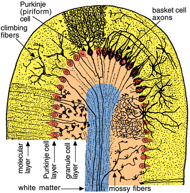
Figure 18—50. Drawing of a transverse section through a cerebellar folium. The folium has a core of white matter (blue) surrounded by cerebellar cortex. The white matter is composed of climbing and mossy corticopedal axons and corticofugal axons from Purkinje (piriform) neurons. The cerebellar cortex consists of three layers (labeled sideways). The molecular layer (yellow) has relatively few cell bodies. The Purkinje (piriform) cell layer is evident as a row of large cell bodies (dark orange). The granule cell layer (pale orange) is composed of small neurons that send axons into the molecular layer. The axons course longitudinally in the folium and synapse on dendritic trees of Purkinje cells. Cell bodies of basket cells (generally located deep in the molecular layer) send axons transversely in the folium. The axons terminate in basket arrangements around adjacent Purkinje cell bodies. (From Cajal, modification of Fig. 200 in Ranson, S. W. and Clark, S. L.: The Anatomy of the Nervous System, 10th ed, Philadelphia, WB Saunders, 1959.)
Go Top