Sulci & Gyri
CLOSE

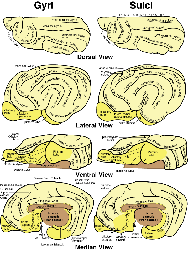
Figure 18—30. Schematic illustration of gyri (left) and sulci (right) of a canine cerebral hemisphere. Dorsal, lateral and ventral views are shown for a left hemisphere; the median view is of a right hemisphere. Neocortex is pale yellow, rhinencephalon is brighter yellow, and major white matter bundles are colored brown. In the piriform lobe of the ventral view, "A" marks the location of the amygdala.
Go Top