Thalamus
CLOSE
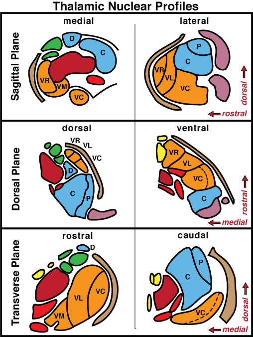
Figure 18—23. Cartoon showing profiles of right-side thalamic nuclei in transverse, dorsal, and sagittal planes of view. Per plane, nuclei are drawn at two levels and directions are shown by arrows. Nuclear groups are color-coded: maroon = dorsomedial nucleus; green = rostral nuclear group; orange = ventral group of lateral nuclei; blue = dorsocaudal group of lateral nuclei; red = intralaminar nuclei; yellow = midline nuclei (in contact with the interthalamic adhesion); brown = reticular thalamic nucleus; and purple = geniculate nuclei (metathalamus). The following abbreviations are used to identify nuclei of the lateral group: C = caudal; D = dorsal; P = pulvinar; VC = ventral caudal; VL = ventral lateral; VM = ventral medial; and VR = ventral rostral. Dotted lines separate medial & lateral pars of the ventral caudal nucleus. Nuclear profiles were copied from: Salazar, I. et al. The Thalamus of the Dog 1989.
Go Top