Pertinent Surgical Anatomy
Lateral Approach to the Femur:
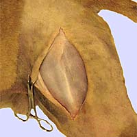
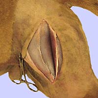
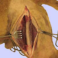
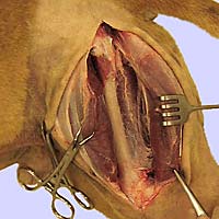
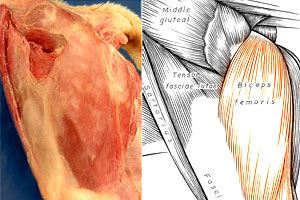
From superficial to deep, a series of cadaver dissections shows the regional anatomy for a lateral approach to repair a fractured femur. (Click to view enlarged images.)
 |
 |
 |
 |
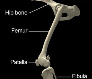
Osteology:
- slight medial curvature of the canine femur results in
compressive forces medially and tensile forces laterally - survey radiographs should always include two views
and the entire hind limb
Musculature:
- the lateral approach to the femur offers great bone
exposure and avoids major vessels and nerves - incising through fascia lata results in minimal bleeding,
also the fascia is easy to suture back together - the adductor muscle attaches to the caudomedial aspect
of the femur and this attachment should be preserved
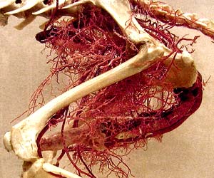
Vasculature:
- major arteries are located medially in the thigh
- rupture of these vessels at the time of trauma can
produce large hematomas
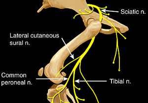
Nerves:
- the sciatic nerve is of major concern because it is the
main nerve supply to the limb - the femoral nerve supplies the quadriceps femoris muscle
and medial cutaneous innetvation
Go Top