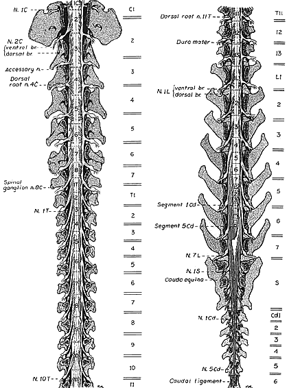
The canine spinal cord is illustrated in situ following removal of vertebral laminae (laminectomy) and removal of dura mater (except along the right side). Locations of intervertebral discs are shown as double lines along the right side.
Spinal cord segments are labeled numerically per region (separated by dotted lines). Except for C1 (which exits via a lateral foramen) cervical spinal nerves exit through intervertebral foramina located cranial to numerically corresponding vertebrae. Because of the presence of a C8 segment (and the absence of a C8 vertebrae), spinal nerves caudal to the cervical region exit through intervertebral foramina formed by caudal margins of numerically corresponding vertebrae.
On the left side of the cervical spinal cord, notice that the spinal root of the accessory nerve originates from the first seven segments of the spinal cord.
Click for more information about relationships between vertebrae and spinal segments and roots for cranial and caudal halves of the spinal cord.