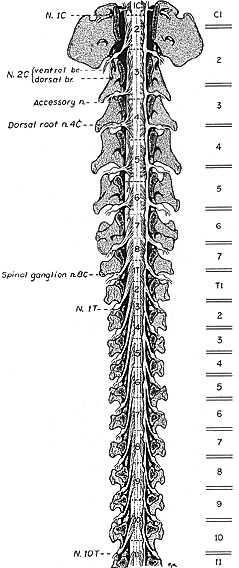
The cranial half of the spinal cord is illustrated in situ following removal of vertebral laminae (laminectomy) and removal of dura mater (except along the right side). Spinal cord segments are labeled numerically per region and separated by dotted lines in this illustration. Locations of intervertebral discs are shown as double lines along the right side.
Relationship: vertebrae and spinal cord segments
Spinal cord segment C3 is the longest segment. Caudal to C3 spinal segments progressively shorten. Caudal to T3 spinal segments progressively lengthen. Spinal cord segments shift position relative to corresponding vertebrae (because length changes in vertebrae are more conservative than those of spinal segments). Shorter segments shift cranially; longer segments shift caudally. It is clinically useful to know that the cervical enlargement (brachial plexus) segments (C6, C7, C8, & T1) are centered over the C6-7 intervertebral disc.
Relationship: vertebrae and spinal roots
Because spinal roots exit at corresponding intervertebral foramina, root length reflects the position of a spinal segment relative to its corresponding vertebra. Segments shifted cranial to their corresponding vertebrae have longer spinal roots.
Because of the presence of a C8 segment (and the absence of a C8 vertebrae), spinal nerves caudal to the cervical region exit through intervertebral foramina formed by caudal margins of numerically corresponding vertebrae.