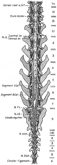
The caudal half of the spinal cord is illustrated in situ following removal of vertebral laminae (laminectomy) and removal of dura mater (except along the right side). Spinal cord segments are labeled numerically per region and separated by dotted lines. Locations of intervertebral discs are shown as double lines along the right side.
Relationship: vertebrae and spinal cord segments
Segments of the thoracolumbar junction are long and located within their corresponding vertebrae. Caudal to the thoracolumbar region, segments progressively shorten. It is clinically useful to know that the sacral segments of the spinal cord lie within the L5 vertebra and the spinal cord ends approximately at the L6-7 vertebral junction. (This relationship applies medium and large adult dogs, for small dogs [< 7 Kg] the spinal cord ends in vertebra S1; it extends even further caudally in the neonatal dog.)
Relationship: vertebrae and spinal roots
Spinal roots exit at intervertebral foramina located at caudal margins of corresponding vertebrae. As spinal segments shorten and shift cranial to their corresponding vertebrae, spinal roots must elongate to reach their target intervertebral foramina. The long spinal roots traveling caudal to the end of the spinal cord (sacral & caudal roots) are collectively known as the cauda equina. Damage to the cauda equina results in clinical signs related to the tail and pelvic viscera.