Lab: Cerebellum
CLOSE
Cerebellar Purkinje Cells
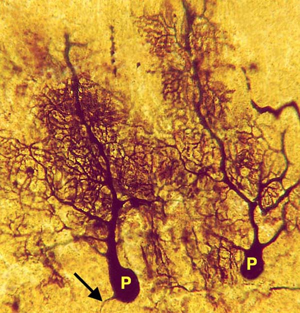
Golgi stain. Two Purkinje cells (P) are coated by silver precipitate. The axon (a) of a Purkinje neuron is evident. For the previous view of Purkinje cells, click here. For a fluorescent view of Purkinje cells, click here. To view the cortical drawing, click here.
Go Top