Lab: Cerebellum
CLOSE
Cerebellar Purkinje Cells
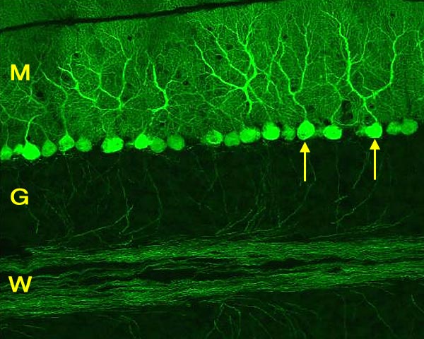
Confocal micrograph from mouse cerebellum showing green-fluorescent protein in Purkinje cells (arrows). The dendrites of Purkinje neurons ascend to the molecular layer (M). The axons of Purkinje neurons pass through the granule cell layer (G) and run in the white matter (W). For the other view of Purkinje cells, click here. To view the cortical drawing, click here.
Go Top