Lab: Cerebellum
CLOSE
Purkinje Cells in Cerebellar Cortex
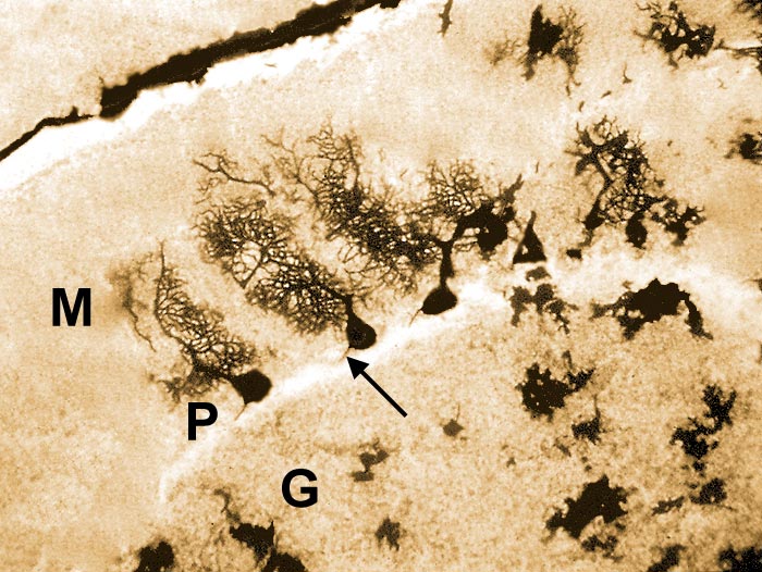
Purkinje cells (neurons) are visible in this pig cerebellar cortex stained by the Golgi method. M = molecular layer, P = layer of Purkinje cell bodies, G = granule cell layer. The arrow points to an axon of a Purkinje neuron. For an enlarged view of Purkinje cells, click here. For a fluorescent view of Purkinje cells, click here. To view the cortical drawing, click here.
Go Top