Brain Anatomy Introduction
CLOSE
Equine Hindbrain
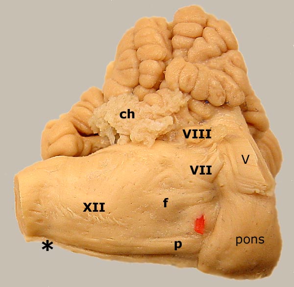
Ventrolateral view of an equine hindbrain. A pyramid (p) emerges form the pons and run along the ventral surface of the myelencephalon. Pyramids disappear from the surface at the juncture with the spinal cord (asterisk), because they turn dorsal and decussate. The red pic is in the trapezoid body. A swelling produced by the motor nucleus of the facial nerve (f) is evident in this specimen. The facial (VII), vestibulocochlear (VIII), and hypoglossal (XII) cranial nerves are labeled. Notice choroid plexus (cp) protruding from the lateral aperture of the fourth ventricle.

Go Top
The trigeminal nerve (V) exits through the pons (white). For another view of pyramids, click here. To return to the sheep image, click here.

Go Top