Brain Anatomy Introduction
CLOSE
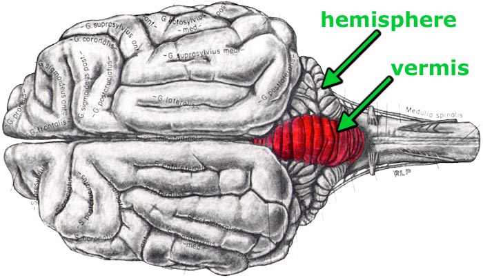
Canine Brain — Dorsal View
Drawing of a canine brain seen from a dorsal view. The vermis of the cerebellum is colored red. Bilateral cerebellar hemispheres flank the vermis. (Note: The term vermis = worm. The central region of the cerebellar appears wormlike? To return to the metencephalon section, click here. To return to the cerebellar nuclei section, click here.
The cruciate sulcus and coronal sulcus along with adjacent gyri are labeled in the cerebrum.
Go Top