Brain Anatomy Introduction
CLOSE
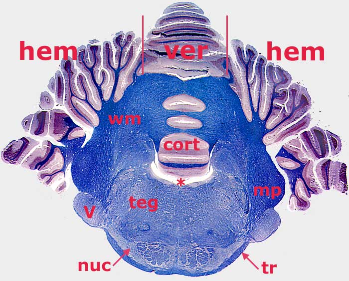
Canine Metencephalon Transection
Transverse section through a canine metencephalon (white matter is stained blue). The cerebellum is dorsal and the pons is ventral. The vermis (ver) of the cerebellum is the middle region; it is flanked by cerebellar hemispheres (hem). Gray matter covering the surface of the cerebellum is cerebellar cortex (cortex). Cerebellar white matter (wm) is deep to the cortex. The middle cerebellar peduncle (mp) is evident in this section. Cerebellar peduncles connect the cerebellum to the brainstem.
The pons consists of a tegmentum (teg) and a distinct ventral part. The latter features pontine nuclei (nuc) which give rise to transverse pontine fibers (tr) that become the middle cerebellar peduncle (mp). The trigeminal nerve (V) penetrates pontine fibers as it exits the metencephalon. An asterisk marks the fourth ventricle, covered by a thin rostral medullary vellum.
To view cerebellar nuclei, click here. For a dorsal view of the cerebellum, click here.
Go Top