Brain Anatomy Introduction
CLOSE
Canine Cerebellum & Medulla Oblongata
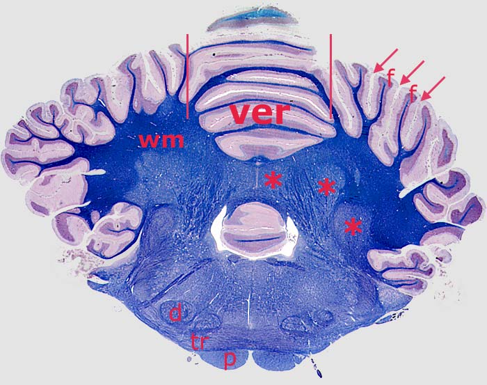
Transverse section through a canine hindbrain (white matter is stained blue). Red lines show approximate sagittal boundaries of the cerebellar vermis (ver). The surface of the cerebellum is arranged into folia (f) separated by sulci (arrows). Cerebellar white matter (wm) is deep to the cerebellar cortex (which appears two-toned). Cerebellar nuclei (asterisks) are embedded in the white matter. To return to the pons section, click here. For a dorsal view of the cerebellum, click here.
Prominent features of the myelencephalon include: pyramid (p), trapezoid body (tr), and dorsal nucleus of the trapezoid body (d).
Go Top