Brain Anatomy Introduction
CLOSE
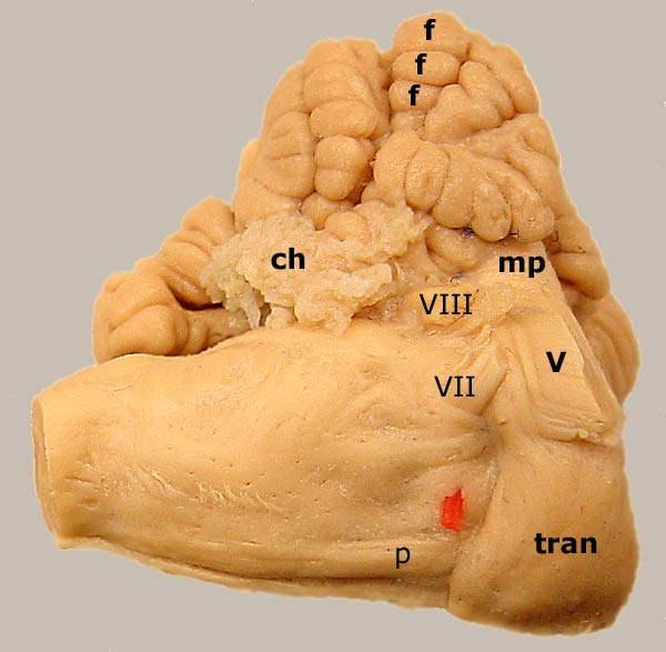
Equine Hindbrain
Ventrolateral view of an equine hindbrain. The cerebellum is dorsal to the pons. Folia (f) of the cerebellar surface are separated by sulci. Ventrally, transverse pontine fibers (tran) mark the boundary of the pons. The transverse pontine fibers form the middle cerebellar peduncle (mp). The trigeminal nerve (V) exits through transverse pontine fibers. To return to the lateral view, click here. For a medial view of the metencephalon, click here.
Labeled myelencephalic structures are: pyramid (p), trapezoid body (red), facial nerve (VII), vestibulocochlear nerve (VIII), and choroid plexus (ch).
Go Top