Brain Anatomy Introduction
CLOSE
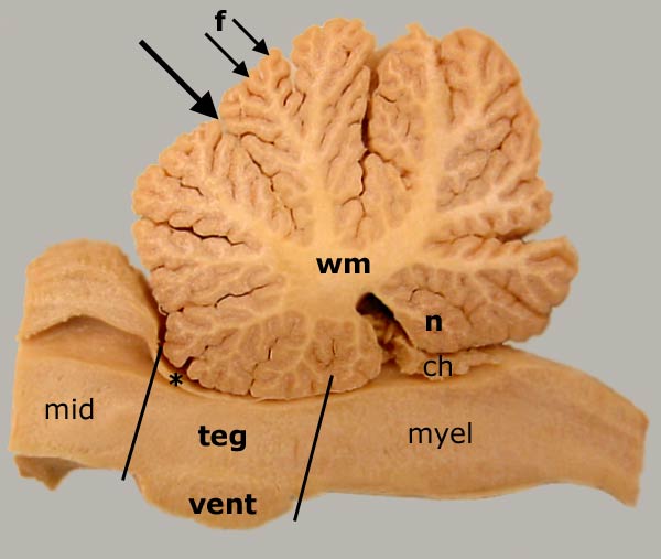
Equine Metencephalon — Medial View
Sagittal section through an equine midbrain and hindbrain. The vermis of cerebellum is section on the midline, exposing cerebellar white matter (wm) deep to cerebellar cortex. Ridges on the surface of the cerebellum are called folia (f, arrows); they are separated by sulci. Fissures divide the vermis into lobules. The most caudal-ventral lobule is the nodulus (n). The large arrow points to the primary fissure (the first to develop embryologically, it divides the cerebellum into rostral and caudal lobes). The pons has a distinct ventral portion (vent) and a dorsal part that is called tegmentum (teg). A rostral medullary vellum (asterisk) forms the metencephalic roof of the fourth ventricle. To return to the lateral view, click here. To see a ventrolateral view, click here.
Other labels indicate midbrain (mid), myelencephalon (myel), and choroid plexus (ch).
Go Top