Brain Anatomy Introduction
CLOSE
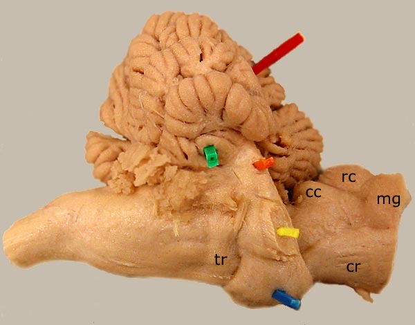
Equine Cerebellum & Pons
Lateral view of an equine cerebellum and caudal brainstem. The green pic is in the flocculus of the cerebellum and the red pic is in the cerebellar primary fissure. Ventrally, transverse pontine fibers (blue) produce a prominent bridge at the surface of the pons. The transverse pontine fibers form the middle cerebellar peduncle (orange). The trigeminal nerve (yellow) exits the brainstem by passing through transverse pontine fibers. For a ventrolateral view, click here. For a medial view of the metencephalon, click here.
The trapezoid body (tr) of the myelencephalon is labeled. Mesencephalic structures are: caudal colliculus (cc), rostral colliculus (rc), and crus cerebri (cr). The medial geniculate body (mg) of the diencephalon is present.
Go Top