Brain Anatomy Introduction
CLOSE
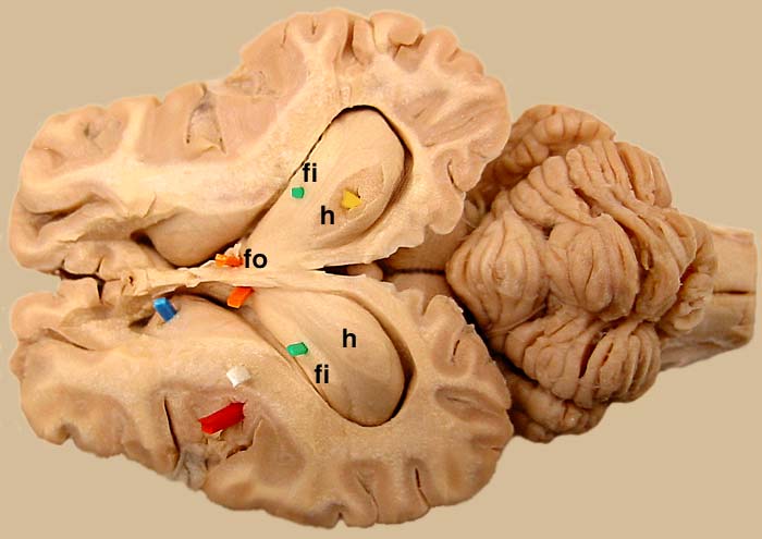
Hippocampus Exposed in Sheep Brain
A sheep brain was dissected to expose the lateral ventricle and the hippocampus (h; yellow) bilaterally. The fimbria (fi; green) appears as a white matter ledge along the lateral margin of the hippocampus. Fibers of the fimbria are continued rostrally as the fornix (fo; orange). To see the hippocampus from a ventral view, click here. To see the hippocampus in more isolation, click here. To return to the original hippocampus page, click here.
Go Top