Brain Anatomy Introduction
CLOSE
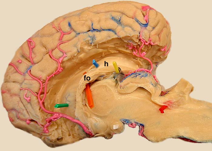
Dissected Equine Half Brain - Medial View
Medial view of an equine half brain. The ventromedial telencephalon has been removed to expose the lateral ventricle. The caudate nucleus (green pic) forms the lateral wall of the ventricle. The floor of the ventricle is formed by the hippocampus (blue; h), which curves caudoventrally. Axons from the hippocampus form the fornix (fo). (The fimbria is not evident because it is positioned lateral to the hippocampus.)
To see a more isolated equine hippocampus, click here.
To see the hippocampus dissected in a sheep brain, click here.
To see the hippocampus in canine brain transections, click here.
To see a more isolated equine hippocampus, click here.
To see the hippocampus dissected in a sheep brain, click here.
To see the hippocampus in canine brain transections, click here.
Go Top