
LAB 19 External Head Dissection
Face and Teeth
Lab Objectives:
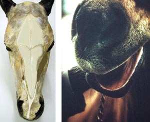
• Start by passing a finger into the nostril to palpate the nasal diverticulum and then pass your finger medially into the nasal cavity and palpate the nasal septum and the ventral nasal meatus. Before splitting the head the skin should be removed in order to observe several midline structures.
• On the dorsal side of the whole head observe the midline tendon of the levator labii superioris muscle and note the left and right tendons that join to form the midline tendon.
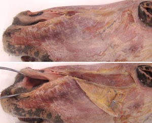
• On the ventral side of the whole head note the sternohyoideus muscles to that insert on the basihyoid bone deep to the large mass of mandibular lymph nodes. Separate these muscles on the midline to expose the ventral aspect of the larynx and trachea.
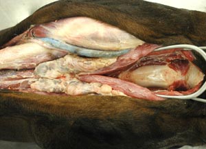
• After splitting the head observe and separate the major facial muscles.
• On the lateral side of the head note the buccal nerves passing over the masseter muscle and the facial artery and vein wrapping around the ventral edge of the masseter along with the parotid duct.
• Deep to the facial muscles find the infraorbital nerve emerging from the infraorbital foramen. Likewise, find the mental nerve emerging from the mental foramen.
• Detach the masseter muscle from the facial crest and reflect it caudally to expose the underlying venous sinuses.
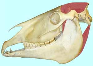
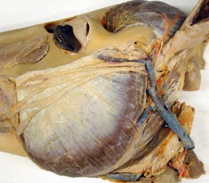
• If time allows, remove the zygomatic arch to expose the eye muscles and the maxillary artery and nerve.
Anatomical Terms:
External Head Structures
nasolabial plate (bov)
orifice of the nasolacrimal duct
nasal diverticulum (eq)
levator labii superioris m. (eq, bov)
infraorbital n.
mandibular lymph nodes
sternohyoideus m.
basihyoid bone
thyroid notch
cricothyroid ligament
thyroid cartilage
masseter m.
dorsal & ventral branches of the facial n.
mental n.
facial vein
venous sinuses of the facial v.
facial a.
parotid salivary gland
parotid duct
post-orbital fat pad
temporalis m.
supraorbital foramen
supraorbital n.
(caudal maxillary sinus)
maxillary a
maxillary n.
medial pterygoid m.
Eye Muscles and Associated Structures
periorbita
lacrimal gland
lateral rectus m.
ventral rectus m.
dorsal rectus m.
ventral oblique m.
retractor bulbi m.
optic n.
Instructor Commentary:
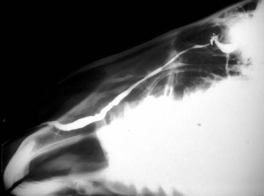
Inside the equine nostril there is a long cutaneous sac known as the nasal diverticulum or false nostril. The nasal diverticulum is caudal to the nostril and is easily palpated by passing a finger into the nostril in a caudal lateral direction so that the finger is close to the skin surface. Medially the finger can be passed into the nasal cavity. When passing a nasogastric tube one should be careful that the tube does not pass into the nasal diverticulum.
The masseter muscle is well developed in herbivores while the temporalis m. is poorly developed. The reverse is true in carnivores and is related to differences in dentition, diet, and lifestyle. Herbivores have large salivary glands in order to moisten great amounts of forage which is often dry. The parotid duct passes over the masseter muscle in the dog but wraps around the ventral edge of the masseter m. in herbivores. Deep to the masseter m. lie several venous sinuses that are compressed by the masseter muscle so that venous blood is pumped from the head into neck veins against gravity when the head is down for grazing.
As the buccal branches of the facial nerve pass over the masseter muscle they are vulnerable to trauma. This can occur when the head of a horse is not well secured during a bumpy trailer ride. Such horses may come out of a trailer with flaccid facial muscles on the side where trauma occurred.
The upper lip anatomy of herbivores varies considerably. It is rigid in cattle but mobile in horse, sheep and goats. The lip curl of equine flehmen behavior occurs due to the action of a midline tendon pulled by left and right levator labii superioris muscles. Sheep and goats have a split upper lip that may facilitate grazing close to the ground.
Dissection Images:
Note: Click an image to see it enlarged, view its caption, and toggle its labels.
| 1 | 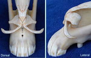 |
 |
2 |
| 3 | 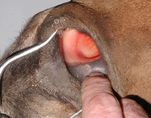 |
 |
4 |
| 5 | 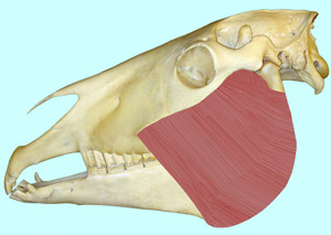 |
 |
6 |
| 7 | 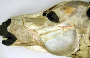 |
 |
8 |
| 9 | 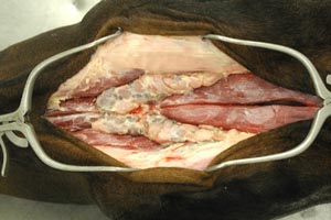 |
 |
10 |
| 11 | 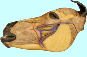 |
 |
12 |
| 13 | 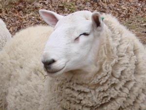 |
 |
14 |
| 15 |  |
 |
16 |
| 17 | 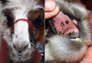 |
 |
18 |