
LAB 20 Internal Head Dissection
Nasal & Oral Cavities, Pharynx & Larynx
Lab Objectives:
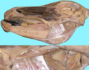
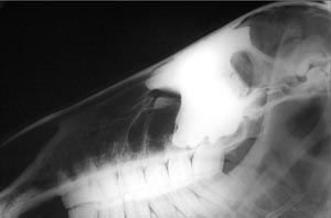
• On the cut midline surface identify the major regions and structures including the nasal cavity, nasopharynx, laryngopharynx, oral cavity and oropharynx. Also note the nasal septum, vomer, hard and soft palates, larynx, and guttural pouches in equine specimens.
• If the nasal septum is present, remove most of it but leave the vomer. Observe the passageways on either side of the vomer. These are the choanae. Observe the dorsal and ventral conchae and the smaller ethmoconchae. Also, identify the large ventral meatus, and the smaller middle meatus and dorsal meatus. Nasogastric tubes must only pass through the ventral meatus to avoid nose bleeds (epistaxis).
• Cut a window in the caudal part of the dorsal concha to expose the conchofrontal sinus and the frontomaxillary opening.
• Find the thin palatopharyngeal folds extending caudally from the caudal end of the equine soft palate. Imagine how these folds and the soft palate form an ostium that surrounds the entrance to the larynx.
• Observe the papillae of the tongue and note species differences. Find the thick palatoglossal folds near the root of the tongue. Caudal to these folds find the palatine tonsils in the horse which lie lateral to the root of the tongue. On bovine specimens, find the orifice of the tonsilar sinus near the ventral lateral surface of the soft palate.
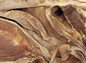
• Ventral to the tongue find the strap like geniohyoid muscles attaching to the basihyoid bone. In this region find the lingual process of the basihyoid bone if the band saw cut left half or more of the basihyoid bone. On the caudal edge of the basihyoid bone find the attachment of the sternohyoid muscles. Dorsal to the geniohyoideus muscles find the genioglossus and hyoglossus muscles forming the extrinsic muscle mass of the tongue.
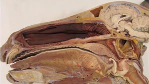
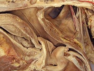
• On the lateral wall of the nasopharynx find the slit like entrance to auditory tube and the guttural pouch. Enlarge the guttural pouch by detachment of the longus capitus muscle from the skull and then reflect the muscle caudally.
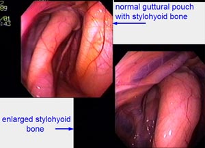
• Adjacent to the thin lining of the guttural pouch note the thin stylohyoid bone that divides the pouch into a small lateral compartment and a larger medial compartment. On the floor of the pouch find the retropharyngeal lymph nodes and look for signs of strangles abscesses. On the walls of the guttural pouch look for internal and external carotid arteries and cranial nerves 9 and 12.
• Identify the laryngeal cartilages, vocal folds and the laryngeal ventricles. Note the ossified rostral ventral part of the thyroid cartilage and the thyroid notch caudal to it. On the dorsal surface of the cricoid cartilage identify the cricoarytenoideus dorsalis muscle.
Anatomical Terms:
Midline Head Structures
nasal septum
dorsal & ventral conchae
middle concha (bov)
ethmoid conchae
conchofrontal space
lingual fissure (bov)
torus linguae (bov)
sublingual caruncles
vallate papillae
geniohyoideus m.
genioglossus m.
hyoglossus m.
palatoglossal fold
palatopharyngeal folds
palatal ridges
buccal papillae (bov)
oropharynx
palatine tonsil
nasopharynx
orifice of the auditory tube
pharyngeal septum (bov)
pharyngeal tonsil (bov)
guttural pouches (eq)
retropharyngeal lymph nodes
longus capitis m.
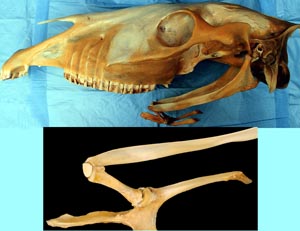
stylohyoid bone
ceratohyoid bone
(basihyoid bone)
thyrohyoid bone
laryngeal cartilages (cricoid, thyroid, arytenoid, epiglottis)
laryngeal ventricle (eq)
vocal folds (eq)
cricoarytenoideus dorsalis m.
cricoarytenoideus lateralis m.
common carotid a.
internal carotid a. (find in eq)
external carotid a.
linguofacial trunk
lingual a.
facial a.
vagosympathetic nerve trunk
cervical sympathetic trunk
cranial cervical ganglion
vagus n.
cranial laryngeal branch
Instructor Commentary:
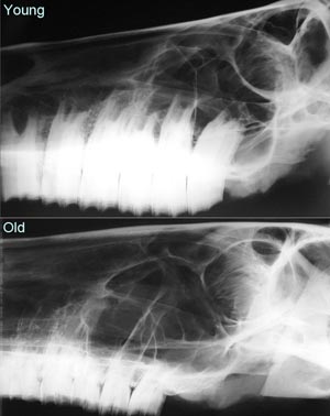
If the midline cut leaves the nasal septum intact, the vomer bone should be found forming the caudal ventral edge of the septum. The vomer marks the place of transition from nasal cavity to the nasopharynx and is referred to as the choana. The vomer separates right and left choanae. Choanal atresia refers to closed or constricted choanae, a defect that is occasionally found in newborn alpacas and Arab foals.
The hard palate separates the nasal cavity from the oral cavity and the soft palate separates the nasopharynx from the oropharynx. The caudal edge of the soft palate is continuous with the palatopharyngeal folds which are best developed in the horse where they appear to wrap around the opening of the larynx and thereby function to seal the laryngeal opening into the caudal end of the nasopharynx during respiration. This tight seal relates to the fact that the horse is an obligate nasal breather.
Roaring is a common term for laryngeal hemiplegia which refers to paralysis (plegia) on one side (hemi) of the larynx. In most cases the larynx is paralyzed on the left side. This predilection can be explained by right/left differences in the path of the recurrent laryngeal nerve in the thorax. On the left side the recurrent laryngeal nerve passes around the ligamentum arteriosum and the aorta but on the right side the recurrent laryngeal nerve loops around the subclavian artery. On the left side the recurrent nerve passes close to the left tracheobronchial lymph nodes but on the right side the recurrent loop is cranial to the tracheobronchial lymph nodes. Damage to the recurrent nerve may be due to spread of inflammatory substances from inflamed left tracheobronchial lymph nodes to the adjacent left recurrent nerve (image 10-4).
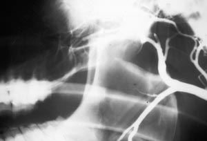
On the lateral walls of the equine nasopharynx there are long slit like openings to the auditory tube but in other species the openings are small and difficult to find. The large openings in the horse relate to the unusual auditory tube which is dilated to form the guttural pouches. The thin lining of the guttural pouches may appear to form a septum between the bilateral pouches depending on how the band saw cut is made. As with the pleural and peritoneal cavities, the guttural pouches are empty but numerous structures can be seen through the thin lining of the pouch. The most prominent structure related to the pouch is the stylohyoid bone which forms a ventral septum that partially divides the pouch into a small lateral compartment and a larger medial compartment. Also seen through the lining of the pouch will be the internal and external carotid arteries, cranial nerves 9 and 12 and medial retropharyngeal lymph nodes which may be abscessed due to a Strep. equi infection known as strangles.
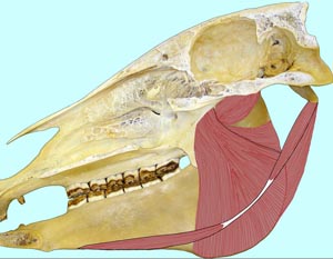
The medial pterygoid muscle lies on the medial side of the mandible in a place similar to that of the masseter m. on the lateral side. Two pairs of muscles consisting of one masseter and the contralateral (opposite side) medial pterygoid muscle act alternately to produce the side to side chewing motions used by horses to grind forage between the cheek teeth.
Dissection Images:
Note: Click an image to see it enlarged, view its caption, and toggle its labels.
| 1 | 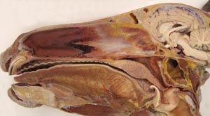 |
 |
2 |
| 3 | 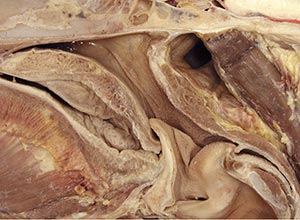 |
 |
4 |
| 5 |  |
 |
6 |
| 7 | 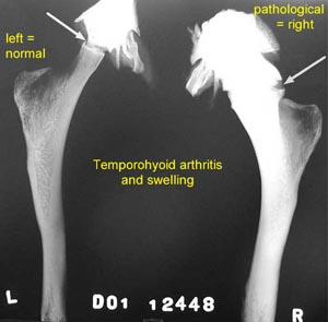 |
 |
8 |
| 9 | 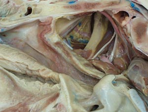 |
 |
10 |
| 11 | 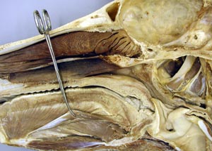 |
 |
12 |
| 13 | 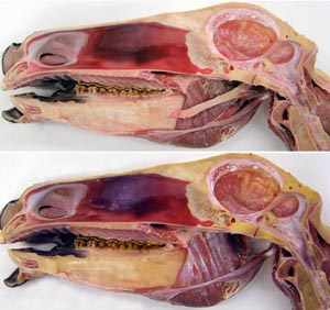 |
 |
14 |
| 15 | 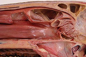 |
 |
16 |
| 17 | 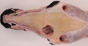 |
 |
18 |
| 19 | 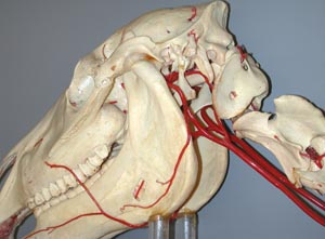 |
 |
20 |
| Previous Lab | Next Lab |