LAB 12 Fistulated Rumen
Explore Bovine Fistulated Rumen
Lab Objectives:
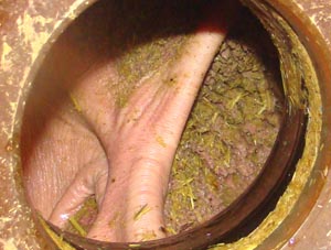
• To observe the dorsal and ventral sacs and associated pillars through a rumen fistula.
• To observe movement of ingesta and rumen structures throughout several contraction cycles.
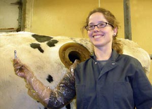
• To palpate rumen and reticular structures and locate a cow magnet in the reticulum. If the fistulated cow is pregnant, to palpate the fetus through the rumen wall.
Anatomical Terms:
Abdominal Wall Muscles and Associated Structures
subiliac lymphocenter (eq)
subiliac lymph node (bov)
external abdominal oblique m.
tunica flava abdominis
rectus abdominis m.
internal abdominal oblique m.
transversus abdominis m.
Equine Stomach and Large Intestines
(equine terms repeat in Lab 13)
cecum
base, body & apex
right & left ventral colon
band
sacculations
pelvic flexure
left & right dorsal colon
small colon
transverse colon
descending colon
Ruminant Stomach Structures
rumen
dorsal, ventral and cranial sacs
dorsal & ventral caudal blind sacs
right & left longitudinal grooves
cranial & caudal pillars
dorsal & ventral coronary pillars
ruminoreticular fold
rumen papillae
reticulum
reticular cells (honeycomb)
reticular groove
cardia (top)
reticulo-omasal orifice (bottom)
omasum
omasal laminae
abomasum
Swine Stomach and Cecum Structures
gastric diverticulum
torus pyloricus
ileal papilla
Camelid Stomach Structures
first compartment
sacculations
transverse fold
second compartment
ventricular lip
third compartment
Instructor Commentary:
A rumen fistula is a specialized gastric fistula. The first gastric fistula was formed in 1822 by an accidental gunshot wound to Alexis St. Martin, a French Canadian voyager. A gastric fistula is useful for study of the digestive process. Food samples can be placed in the rumen and then removed periodically for analysis to determine the rate and degree of digestion
The rumen is the easiest part of the bovine stomach to fistulate because most of the stomach is hidden by the rib cage. The caudal dorsal part of the rumen lies deep to the left paralumbar fossa where the abdominal wall is thin and does not support the viscera. Additionally, the stratified squamous lining of the rumen is tougher than the glandular epithelium of the abomasum. When making a rumen fistula, the rumen wall is first sutured to the body wall in the paralumbar region to create a tight seal that will prevent infection from entering the peritoneal cavity. After this seal is made, the rumen is opened and the edges of the ruminal incision are sutured to the skin.
The horizontal cranial and caudal pillars mark the boundary between the dorsal and ventral ruminal sacs. A third sac, the cranial sac lies cranial to the cranial pillar. The cranial sac is also known as the atrium ruminis because food (ingesta) enters the rumen through the atrium ruminis. Cranial to the atrium ruminis is the small reticular compartment that is separated from the rumen by a thin U shaped wall, the ruminoreticular fold. The reticulum and rumen form a functional ruminoreticular compartment. Contraction of the walls of the ruminoreticulum cause mixing of the semifluid ingesta and thereby enhance the fermentative process. Typically there are about two contractions per minute causing the fluid level to rise and fall.
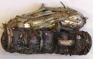
Cow magnets are put in the reticulum of dairy cows by manual placement of the magnet in the throat so that the cow is forced to swallow it. The cow magnet prevents sharp objects in the feed from causing penetration of the stomach wall. Historically "hardware disease" resulted from foreign objects that entered the feed from junk laden barnyards and fields. Modern mechanical grinding equipment used to provide TMR (total mixed rations) feeding can introduce metal filings into the feed.
Dissection Images:
Note: Click an image to see it enlarged, view its caption, and toggle its labels.
| 1 | 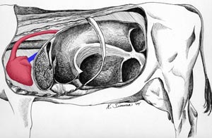 |
 |
2 |
| 3 | 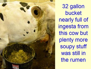 |
 |
4 |
| 5 | 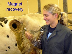 |
 |
6 |
