LAB 13 Equine Abdominal Viscera
Identify and Remove Abdominal Viscera
Lab Objectives:
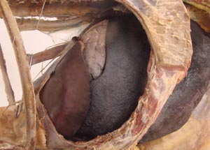
• To observe the stomach, spleen and liver through windows cut in the diaphragm.
• To observe the spleen and nephrosplenic ligament in the left dorsal paralumbar region.
• To observe the base of the cecum and caudal loop of the duodenum in the right dorsal paralumbar region.
• To identify as many abdominal structures as possible by palpation from the dorsal paralumbar region on both sides. This will include genital structures in the pelvic inlet.
• To observe more and more of the abdominal viscera as the body wall is progressively reflected from dorsal to ventral. To find the epiploic foramen on the right side ventral to the liver.
• To remove all support of the viscera by the body wall and then note visceral attachments to various structures near the dorsal midline.
• Remove the abdominal viscera as a single unit including the abdominal aorta, and diaphragmatic crura.
Anatomical Terms:
Equine Abdominal Structures
pelvic brim
ductus deferens
vaginal ring (male)
(terms sequence follows demo viscera)
left kidney
spleen
base
apex
nephrosplenic space
nephrosplenic ligament
epiploic foramen
caudal vena cava
hepatic veins
liver
left, right, quadrate, & caudate lobes
portal v.
bile duct
esophagus
stomach
cardia
fundus
pyloric region
pylorus
lesser curvature
lesser omentum
greater curvature
glandular & non-glandular regions
margo plicatus
duodenum
descending duodenum
major duodenal papilla
caudal flexure/caudal loop
mesoduodenum
jejunum
mesojejunum
root of the mesentery/mesenteric root
ileum
ileocecal fold
cecum
base, body & apex
ileal orifice
cecocolic orifice
cecocolic fold
right & left ventral colon
ventral diaphragmatic flexure
band
sacculations
pelvic flexure
left & right dorsal colon
dorsal diaphragmatic flexure
mesocolon
small colon
transverse colon
descending colon
antimesenteric band
mesenteric band
Equine Abdominal Arteries
(aorta)
celiac a.
splenic a.
hepatic a.
left gastric a.
cranial mesenteric a.
jejunal aa.
right colic aa.
Instructor Commentary:
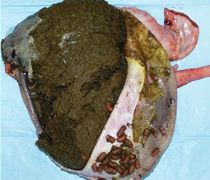
The equid gut has a small stomach, relatively short small intestine and a voluminous large intestine. The large intestine has 3 parts, cecum, large colon, and small colon. All three parts are characterized by longitudinal bands and transverse infolding of the intestinal wall to from serial sacs known as sacculations. The longitudinal bands are formed by longitudinal smooth muscle fibers that are concentrated to form bands rather than being evenly spread over the intestinal surface. The cecum and parts of the large colon have three or four bands while the small colon has only two bands. Some of the bands are hidden in the attached mesocolon but are easily palpated. Therefore, the bands can be referred to as free bands vs. mesocolic bands. The function of the bands is to pull the wall into pleats or infoldings similar to pleats in cloth as with drapery. The infoldings form a series of sacculations. Alternate contraction and relaxation of the circular muscle of the sacculations causes mixing of the ingesta in a side to center fashion. The sacculations are a.k.a haustra and the mixing action is referred to as haustral churning. Bands and sacculations are characteristic of the large intestine of horse, pig and human but are not found in carnivores and ruminants. In the pig the sacculations are more subtle than those of the horse. In humans inflammation of colonic sacculations results in a painful condition known as diverticulitis.
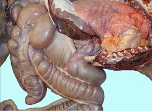
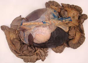
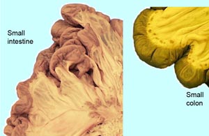
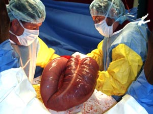
The large colon has two U shaped loops, the ventral and dorsal colons which are joined on the left side at the pelvic flexure which lies near the pelvic inlet. The ventral colon has uniform diameter and 4 bands on both sides. In contrast, the left dorsal colon has only one band, lacks sacculations and has a small diameter. The right dorsal colon has a large diameter, 3 bands and subtle sacculations. The cecum is comma shaped with a wide base in the right dorsal paralumbar region. The apex of the cecum lies between the left and right ventral colons. The small colon is readily distinguished from the small intestine by sacculations, a prominent antimesenteric band and larger diameter. The equid small colon corresponds to the descending colon of other species but it is elongated.
On the left side of the cecal base the ileum enters the cecum via the ileocecal orifice. Nearby this orifice is the cecocolic orifice where cecal contents pass into the right ventral colon.
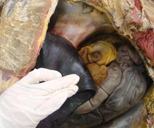
The equine spleen is large and triangular with a wide and thick base dorsally and a thinner apex ventrally. Use of barbiturate anesthesia for euthanasia causes enlargement of the spleen due engorgement with blood. Therefore, the spleen may be abnormally large in cadaver specimens. In life, the spleen stores blood that can be injected into the circulation to increase oxygen carrying capacity during exertion. The large spleen of equids is a reflection of their athletic potential.
The base of the spleen attaches to the left kidney by way of a nephrosplenic ligament. Dorsal to this ligament is the nephrosplenic space between the spleen and the left kidney. Displacement of the left colons into the nephrosplenic space is a common cause of equine colic. Another cause of colic is displacement of a loop of the small intestine into the epiploic foramen. Small bowel entrapment in the epiploic foramen has been associated with the vice known as cribbing (AAEP proceedings 49:371-372; 2003). Strangulation of the gut by a pedunculated lipoma arising from the mesojejunum is another possible cause of colic.
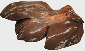
Equids, and many other herbivores, lack a gallbladder but the quadrate lobe can be identified by noting the midventral falciform ligament which is a remnant of the umbilical vein. The quadrate lobe of the liver is adjacent to this ligament on the right side.
Dissection Images:
Note: Click an image to see it enlarged, view its caption, and toggle its labels.
| 1 | 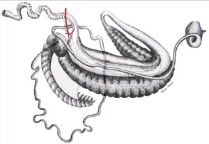 |
 |
2 |
| 3 | 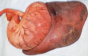 |
 |
4 |
| 5 | 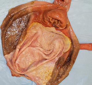 |
 |
6 |
| 7 | 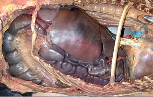 |
 |
8 |
| 9 | 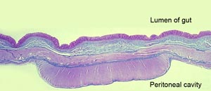 |
 |
10 |
| 11 | 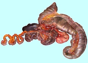 |
 |
12 |
| 13 | 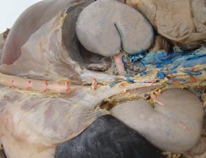 |
 |
14 |
| 15 | 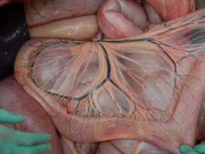 |
 |
16 |
| 17 |  |
 |
18 |
