LAB 11 Abdominal Wall
Abdominal Wall Muscles
Lab Objectives:
• To expose the external abdominal oblique muscle and observe the elastic tunic and the muscle tendon junction.
• To cut the external oblique muscle near its origins on the ribs and reflect the muscle ventrally to expose the internal abdominal oblique muscle.
• To reflect the dorsal part of the internal abdominal oblique muscle to expose the transversus abdominis muscle.
Anatomical Terms:
Abdominal Wall Muscles and Associated Structures
subiliac lymphocenter (eq)
subiliac lymph node (bov)
external abdominal oblique m.
tunica flava abdominis
rectus abdominis m.
internal abdominal oblique m.
transversus abdominis m.
Equine Stomach and Large Intestines
(equine terms repeat in Lab 13)
cecum
base, body & apex
right & left ventral colon
band
sacculations
pelvic flexure
left & right dorsal colon
small colon
transverse colon
descending colon
Ruminant Stomach Structures
rumen
dorsal, ventral and cranial sacs
dorsal & ventral caudal blind sacs
right & left longitudinal grooves
cranial & caudal pillars
dorsal & ventral coronary pillars
ruminoreticular fold
rumen papillae
reticulum
reticular cells (honeycomb)
reticular groove
cardia (top)
reticulo-omasal orifice (bottom)
omasum
omasal laminae
abomasum
Swine Stomach and Cecum Structures
gastric diverticulum
torus pyloricus
ileal papilla
Camelid Stomach Structures
first compartment
sacculations
transverse fold
second compartment
ventricular lip
third compartment
Instructor Commentary:
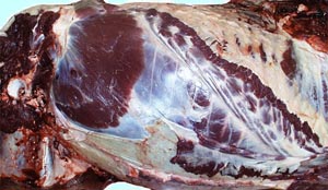
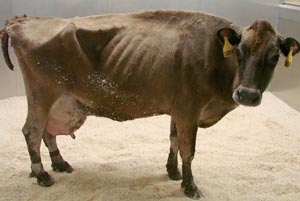
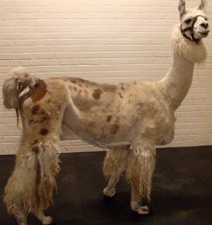
The abdominal cavity of herbivores is large due to a larger gut than is found in carnivores. This larger gut is heavy and thus the abdominal wall must be strong to support the gut. The gut is larger in ruminants than equids and therefore the abdominal cavity of ruminants is deeper and more voluminous. With a heavier gut in ruminants, a stronger abdominal wall would be expected in ruminants when compared with equids. However, the abdominal wall is stronger in equids and therefore, parturition (birthing) in horses is much faster, and sometimes violent. The paralumbar region of the body wall lies between the last rib and the pelvis. As shown in image 1 below, the paralumbar region of the body wall is much larger in ruminants and therefore it is a useful place for surgical entry into the abdominal cavity in a standing animal. The paralumbar region is large enough to pull out a calf or lamb but not large enough in a mare to deliver a foal by C-section.
Besides being large in ruminants, the paralumbar region is a favorable surgical site due to the thin abdominal wall in the dorsal part of this region. The ruminant abdominal muscles are so thin here that a fossa is present caudal to the last rib. The paralumbar fossa is characteristic of ruminants but is absent in equids. The triangular boundaries of the paralumbar fossa are the last rib, the lateral edge of the lumbar transverse processes and the dorsal edge of the thick part of the internal abdominal oblique muscle. This muscle is thin dorsally but thicker ventrally where it supports the viscera. The thick dorsal edge of the internal oblique muscle extends from the tuber coxae to the lower half of the last rib.
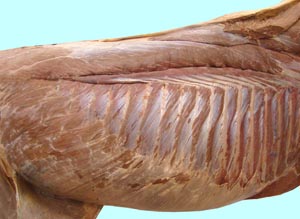
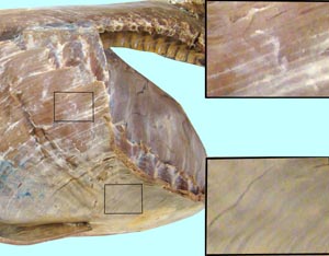
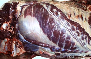
The external abdominal oblique muscle is the strongest of the three lateral abdominal muscles. It appears to be more extensive in the pony than in the horse (compare images 2 & 3 below). The external oblique muscle originates from the ribs while the internal oblique muscle mainly originates from the coxal tuber. In some types of pulmonary disease, the abdominal muscles hypertrophy in order to aid expiration. Hypertrophy of the external abdominal oblique muscle results in a prominent difference in thickness of the muscle and tendinous parts of the external oblique muscle. This thickening results in an observable line along the muscle tendon junction. Heaves is a lay term for forced expiration. Therefore, the exaggerated muscle tendon junction of the external oblique muscle is referred to as a heave line. The ventral part of the external oblique muscle is covered with an elastic deep fascia known as the tunica flava (yellow) abdominis.
Dissection Images:
Note: Click an image to see it enlarged, view its caption, and toggle its labels.
| 1 | 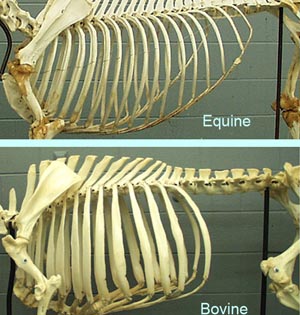 |
 |
2 |
| 3 | 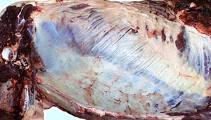 |
 |
4 |
| 5 |  |
 |
6 |
| 7 | 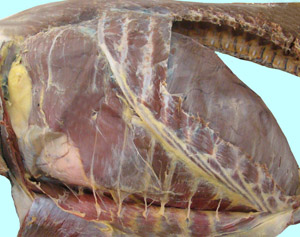 |
 |
8 |
| 9 | 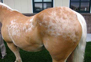 |
 |
10 |
| 11 | 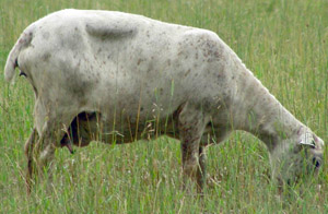 |
 |
12 |
