Brain Anatomy Introduction
CLOSE
Canine and Equine Brainstems
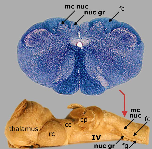
Top: Transection through the the brainstem of a dog at the juncture with the spinal cord. The nucleus gracilis (nuc gr), medial cuneate nucleus (mc nuc), and fasciculus cuneatus (fc) are labeled.
Bottom: Dorsal view of a half brainstem from a horse. The red arrow shows the level of transection for the top image. Locations of the nucleus gracilis (nuc gr), fasciculus gracilis (fg), medial cuneate nucleus (mc nuc), fasciculus cuneatus (fc), and floor of the fourth ventricle (IV) are visible. Also labeled are: cerebellar peduncles (cp), caudal colliculus (cc), rostral colliculus (rc), and thalamus.
To return to the sheep brainstem, click here. To see a drawing of a canine brainstem, click here.
Go Top