Brain Anatomy Introduction
CLOSE
Sheep Brainstem — Dorsal View
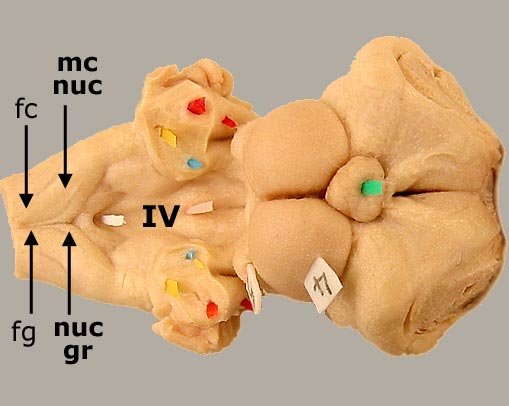
Dorsal view of a sheep brainstem. The fasciculus gracilis (fg) terminates in the nucleus gracilis (nuc gr). The fasciculus cuneatus (fc) coveys axons to the medial cuneate nucleus (mc nuc). The floor of the fourth ventricle (IV) is marked white & pink pics. To see a drawing of a canine brainstem, click here. To see the labeled nuclei in transection, click here.
Also marked: cerebellar peduncles (rostral = blue; middle = red; caudal = yellow), rostral colliculus (4), and pineal body (green).
Go Top