Canine Brainstem — Dorsal View
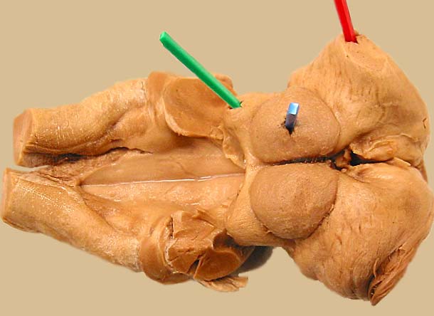
The cerebellum has been removed to expose the dorsal surface of the myelencephalon (the caudal half has been torn). Vestibular nuclei are evident in the floor of the foruth ventricle, medial & caudal to the cut cerebellar peduncles. Nucleus gracilis & cuneate nuclei are evident in the lateral wall at the brain-spinal junction. (Pics are inserted into the caudal colliculus (green), rostral colliculus (blue), and lateral geniculate (red). Click here, for another view.
LABELS
Go Top
RETURN