
LAB 12 Introduction
Thoracic Cavity:
Autonomic Nerves and Heart
(Guide to the Dissection of the Dog, 8th ed., pp. 119-125)
CONTENTS:
Lab Objectives:
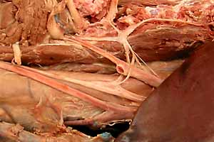
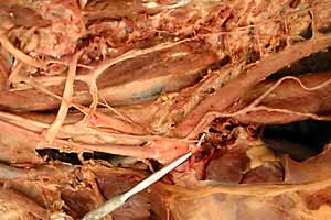
• Examine autonomic (visceral efferent) nerves of the thoracic cavity and identify them as:
- sympathetic (most of the nerves and all of the ganglia in the thoracic cavity)
- parasympathetic (the vagus nerve and its visceral branches)
- sympathetic & parasympathetic combined (vagosympathetic nerve trunk)
• Identify somatic efferent branches of the vagus nerve:
- right recurrent laryngeal n. (loops around subclavian a.)
- left recurrent laryngeal n. (loops around ligametum arteriosum)
(also, the phrenic n. is somatic efferent in fiber type)
• Identify serous membrane and endothoracic fascia tissues surrounding the heart.
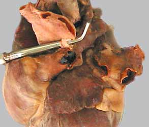
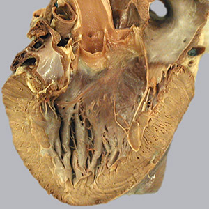
• Dissect the heart to learn its anatomy and develop an understanding of blood flow through the
four chambers of the heart.
Anatomical Terms:
Nerves and ganglia (the following nerves are autonomic & bilateral)
sympathetic trunk
cervicothoracic ganglion
vertebral nerve
ansa subclavia
middle cervical ganglion
cardiac nerves
vagosympathetic trunk
cranial cervical ganglion (not visible, will be dissected in Lab 25)
vagus nerve
recurrent laryngeal nerve
caudal laryngeal nerve
dorsal & ventral vagal branches
dorsal & ventral vagal trunks
Heart and pericardium
(Know: valve locations in a live animal & blood flow though the heart)
fibrous pericardium (fibrous wall of the pericardial cavity/sac)
phrenicopericardial ligament
serous pericardium (lines the pericardial cavity/sac)
parietal pericardium
visceral pericardium (epicardium)
Note: the wall of the pericardial cavity/sac consists physically of three inseparable layers:
pericardial mediastinal pleura
fibrous pericardium &
parietal (serous) pericardium )
heart surfaces: auricular (left) & atrial (right )
coronary groove
subsinuosal interventricular groove
paraconal interventricular groove
right atrium
sinus venarum, contains the following four openings:
caudal vena cava
cranial vena cava
coronary sinus
right atrioventricular orifice
interatrial septum
intervenous tubercle
fossa ovalis
crista terminalis
right auricle
pectinate muscles
endocardium (lining of the heart)
right atrioventricular orifice
right atrioventricular valve (human tricuspid valve)
(parietal & septal cusps)
right ventricle
chordae tendineae
papillary muscles
trabeculae carneae
trabecula septomarginalis (moderator band)
conus arteriosus
pulmonary valve (three semilunar cusps)
pulmonary trunk (splits into right & left pulmonary aa.)
ligamentum arteriosum (fetal ductus arteriosus; connection to the aorta)
Note: pulmonary vv. from the lungs enter the left atrium:
left atrium
left auricle
left atrioventricular orifice
left atrioventricular valve (human mitral/bicuspid valve)
(parietal & septal cusps)
left ventricle
aortic valve (three semilunar cusps)
right coronary artery
left coronary artery
circumflex branch
paraconal interventricular branch
great cardiac vein (drains into the coronary sinus)
Note:
autonomic [Greek: auto = self; nomos = rule/govern] = self governing
Instructor Commentary:
The nerves found in the thoracic cavity are either sympathetic or parasympathetic autonomic nerves, with two exceptions. The recurrent laryngeal nerve carries somatic efferent axons to the larynx while the phrenic nerve carries somatic efferent fibers to the diaphragm.
The connection between the vagus nerve and the pharyngeal arches that give rise to the larynx is established early in development. As the heart migrates from the region of the pharynx to the thoracic cavity, it pulls nerves with it, causing the laryngeal nerve supply to detour into the thoracic cavity on its way to the larynx.
Because of right/left differences in vessel formation, the recurrent laryngeal nerve goes around a different vessels on the right side (subclavian a.) than on the left side (ligamentum arteriosum). The recurrent laryngeal nerve is named caudal laryngeal nerve in the vicinity of the larynx.
The heart is more easily studied in ungulates because the large animal hearts are larger and not filled with latex. Learn what you can now and reinforce that knowledge later in the course
You may choose to read pages 109-115 in Guide to the Dissection of the Dog regarding the Autonomic Nervous System.
Also, when you are on-line, you could click this link to explore the courseware web site: Canine Autonomic Nervous System Pathways (http://vanat.cvm.umn.edu/ans/).
Dissection Steps:
Click to view a PDF list of dissection procedures for this lab:
Show List of Dissection Steps (PDF)
Dissection Images:
Note: Click an image to see it enlarged, view its caption, and toggle its labels.
| 1 | 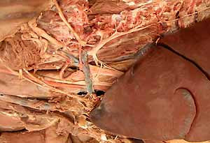 |
 |
2 |
| 3 | 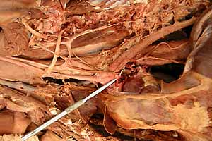 |
 |
4 |
| 5 |  |
 |
6 |
| 7 |  |
 |
8 |
| 9 | 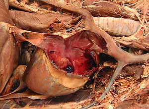 |
 |
10 |
| 11 | 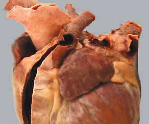 |
 |
12 |