
LAB 11 Introduction
Thoracic Cavity:
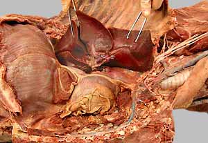
Lungs and Major Vessels
(Guide to the Dissection of the Dog, 8th ed., pp. 107-115)
CONTENTS:
Lab Objectives:
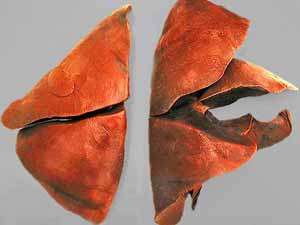
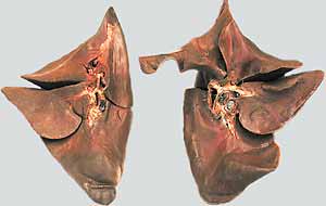
• Identify the lobes of the lungs:
Left Lung
Right Lung
cranial lobe - cranial part
cranial lobe
cranial lobe - caudal part
middle lobe
caudal lobe
caudal lobe
accessory lobe
• Find:
- cardiac notch (between cranial & middle lobes of right lung)
- tracheobronchial lymph nodes (between principal bronchi)
- accessory lobe of the right lung (tucked within a pocket formed by plica vena cava)
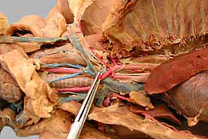
• Veins:
- caudal vena cava
- azygos v.
- cranial vena cava
brachiocephalic v.
external jugular v.
subclavian v.
• Arteries:
- ascending aorta
(left & right coronary aa. will be seen later)
- aortic arch
brachiocephalic trunk
left & right common carotid aa.
right subclavian a.
left subclavian a.
vertebral a.
costocervical trunk
internal thoracic a.
superficial cervical a.
(the subclavian a. becomes axillary a. lateral to ribs)
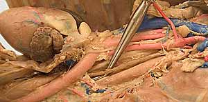
- descending aorta
intercostals aa.
bronchoesophageal a.
• Also:
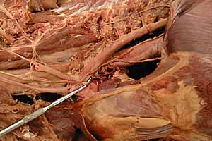
- identify phrenic n. bilaterally
- look for the thoracic duct between the aorta & the azygos v. (the cisterna chyla will be seen later)
Anatomical Terms:
The Lungs
left lung
cranial lobe (cranial & caudal parts)
caudal lobe
aortic impression
right lung
cranial lobe
cardiac notch
middle lobe
caudal lobe
accessory lobe
principal (mainstem) bronchi
carina
lobar bronchi
tracheobronchial lymph nodes
Thorax: vessels cranial to the heart
cranial vena cava
brachiocephalic vein (right & left)
external jugular vein (right & left)
subclavian vein (right & left)
azygos vein
thoracic duct
cisterna chyli (within abdomen, no need to dissect)
tracheal lymph ducts (run in the carotid sheath)
aorta:
ascending aorta
aortic arch
descending aorta
coronary arteries (right & left; will find in Lab 12)
brachiocephalic trunk
left common carotid a.
right common carotid a.
right subclavian a.
left subclavian a. (both right & left subclavian aa. give rise to the following four branches)
vertebral artery
costocervical trunk
superficial cervical a.
internal thoracic a.
axillary a. (continuation of each subclavian a. lateral to the first rib)
Thoracic aorta: branches
(dorsal) intercostal arteries (right & left)
multiple branches to esophagus & bronchi (bronchoesophageal aa.)
phrenic nerve
Note:
azygos = unpaired, from a = not & zygon [Greek] = pair or yoke
Instructor Commentary:
The number of lobes per lung is based on the pattern of lobar bronchi that branch from the principal bronchus, rather than the apparent lobation. Thus the left lung has a cranial lobe with two parts (based on lobar bronchi) rather than having cranial and middle lobes as defined for the right lung.
The term "root" of the lung refers collectively to the vessels and lobar bronchi that attach the lung to the mediastinum. The term "hilus" refers to the region of the lung where components of the root can be seen entering the lung.
Pulmonary arteries usually contain blue latex; pulmonary veins may contain red latex. (Latex does not pass through lung capillaries.):
Blue latex was injected into the external jugular vein of the cadaver. Given enough latex under sufficient pressure, the normal flow is right atrium, right ventricle, pulmonary trunk and pulmonary arteries.
Red latex was injected into the common carotid a. of the cadaver. The latex naturally fills the aorta. To enter the left ventricle, red latex must rupture the aortic valve. To enter the left atrium, red latex must rupture the left atrioventricular valve. Once in the left atrium red latex naturally fills pulmonary veins.
Dissection Steps:
Click to view a PDF list of dissection procedures for this lab:
Show List of Dissection Steps (PDF)
Dissection Images:
Note: Click an image to see it enlarged, view its caption, and toggle its labels.
| 1 |  |
 |
2 |
| 3 | 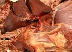 |
 |
4 |
| 5 | 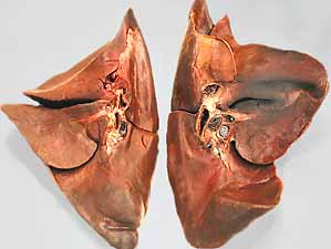 |
 |
6 |
| 7 | 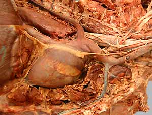 |
 |
8 |
| 9 |  |
 |
10 |
| 11 | 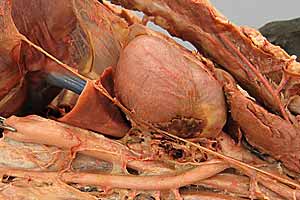 |
 |
12 |