
LAB 8 Introduction
Hypaxial Muscles:
Neck, Thorax, and Abdomen
(Guide to the Dissection of the Dog, 8th ed., pp. 84-90)
CONTENTS:
Lab Objectives:
• Remove superficial fascia from the neck and trunk, as necessary. Identify and preserve
thoracolumbar deep fascia.
•Locate two remaining hypaxial muscles of the neck (longus colli m. and longus capitis m.).
[Previously, sternocephalicus, sternothyroideus, and sternohyoideus mm. were identified.]
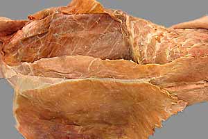
• Identify the thoracic wall muscles involved in breathing (inspiration and expiration).
Inspiratory muscles:
- scalenus m. (three parts)
- serratus dorsalis cranialis m.
- external intercostal mm. (12) [when they pull curved ribs cranially]
Expiratory muscles:
- serratus dorsalis caudalis m. (three small pieces)
- internal intercostal mm. (12) [when they pull curved ribs caudally]
- external abdominal oblique m. (mainly an abdominal wall muscle)
(Also, on the thoracic wall: serratus ventralis m. and rectus thoracis m.)
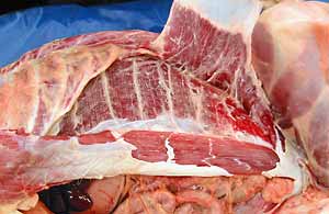
• Identify the four muscles of the abdominal wall:
- external abdominal oblique m.
- internal abdominal oblique m.
- transversus abdominis m.
- rectus abdominis m.
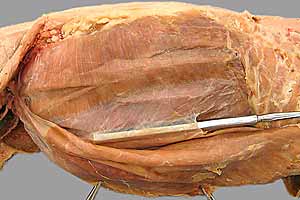
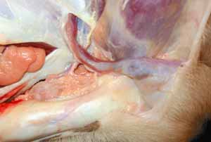
• Observe features of the inguinal canal:
- superficial inguinal ring (in the aponeurosis of the external abdominal oblique m.)
- deep inguinal ring (a triangular gap with three boundaries: inguinal ligament,
rectus abdominis m., & internal abdominal oblique m.)
Anatomical Terms:
Trunk and Neck: hypaxial muscles, etc.
longus capitis m.
longus colli m.
scalenus m.
serratus ventralis m. (cervicis & thoracis)
serratus dorsalis mm. (cranialis & caudalis)
external intercostal m.
internal intercostal m.
Abdominal Wall
linea alba [palpate]
external abdominal oblique m.
inguinal ligament (along the thigh, adjacent to the vascular lacuna)
internal abdominal oblique m.
cremaster m. (male dog; usually absent in cats, which have a levator scroti m.)
transversus abdominis m.
rectus abdominis m. [palpate]
Inguinal Canal
superficial inguinal ring
deep inguinal ring
vaginal process (female)
vaginal tunics (of vaginal process) covering spermatic cord (male)
Note:
alba [Latin] = white
serratus [Latin] = serrated (from serra [L.] = saw)
scalenus [Latin & Greek] = uneven
inguinal [Latin] = pertaining to the groin
tunica [Latin] = covering or coat
vagina [Latin] = sheath
Instructor Commentary:
Based on location, embryonic origin, and innervation, muscles of the trunk and neck can be categorized as epaxial or hypaxial. Hypaxial (hypo-axial) muscles are generally located below the spine (ventral to vertebral transverse processes), although some hypaxial muscles extend dorsal to the spine. A better criterion is that hypaxial muscles are innervated by ventral branches of spinal nerves. (In contrast, epaxial muscles are located dorsal to the vertebral column and are innervated by dorsal branches of spinal nerves.)
Breathing involves inspiration (expanding the chest wall and thoracic cavity volume) and expiration (collapsing the wall and decreasing the volume). Although the diaphragm is the principal muscle affecting thoracic cavity volume, muscles of the thoracic wall also play a role, particularly during maximal exertion. Because ribs are curved, muscles that pull them forward expand the thoracic cavity. In contrast, muscles of expiration act to rotate ribs caudally.
Anatomically, the linea alba (white line) is a raphe, a seam formed by bilateral muscles attaching to one another in regions where bone is absent. Surgically, the linea alba is the preferred site for entering the carnivore abdomen because it is bloodless and easily sutured.
The terms inguinal canal and inguinal ligament refer to defined entities rather than a distinct physical canal or ligament. The inguinal canal is just gaps in each of two adjacent layers of body wall, through which structures pass from inside to outside the abdomen. The inguinal ligament is the caudal edge of the aponeurosis of the external abdominal oblique m., but it is not particularly thickened in the carnivore.
Dissection Steps:
Click to view a PDF list of dissection procedures for this lab:
Show List of Dissection Steps (PDF)
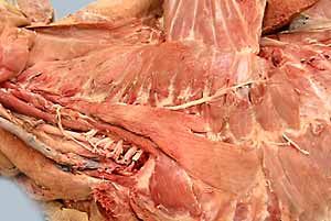
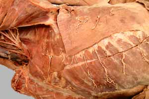
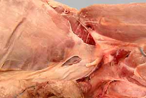
Dissection Images:
Note: Click an image to see it enlarged, view its caption, and toggle its labels.
| 1 | 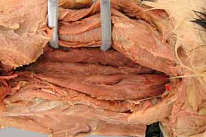 |
 |
2 |
| 3 | 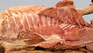 |
 |
4 |
| 5 | 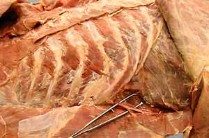 |
 |
6 |
| 7 | 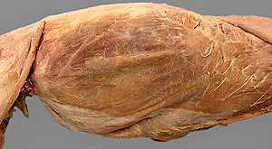 |
 |
8 |
| 9 | 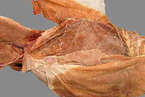 |
 |
10 |
| 11 | 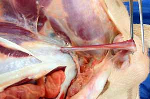 |
 |
12 |
| 13 | 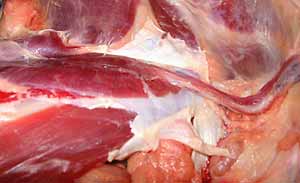 |
 |
14 |