
LAB 7 Introduction
Pelvic Limb Muscles:
Caudal Crus and Hip & Stifle Joints
(Guide to the Dissection of the Dog, 8th ed., pp. 68-77)
CONTENTS:
Lab Objectives:
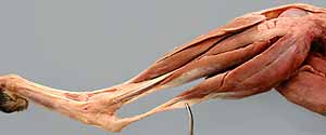
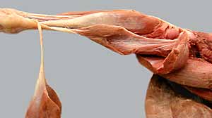
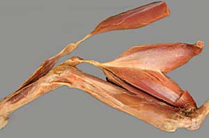
• Dissect the four (six in the cat) major muscles comprising caudal muscles of the crus (leg):
- gastrocnemius m. (medial and lateral heads)
- superficial digital flexor m. (has a calcanean bursa)
- deep digital flexor m. (lateral and medial heads)
- popliteus m.
and in the cat:
- soleus m., a third head of the cat gastrocnemius m. (absent in dogs)
- caudal tibial m., positioned between lateral & medial heads of deep digital flexor m.
•Identify components of the common calcanean tendon
• Examine pelvic limb joints:
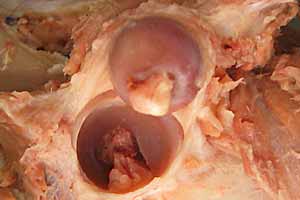
- sacroiliac joint
- pelvic symphysis
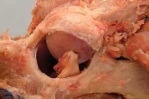
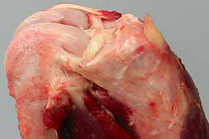
- hip joint (ligament of head of femur & transverse acetabular ligament)
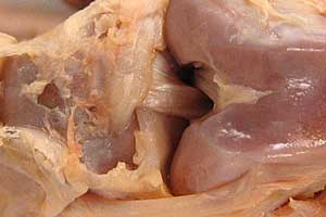
- stifle joint (collateral, cruciate, & femoropatellar ligaments; and
bilateral menisci)
Anatomical Terms:
Pelvic Limb: muscles & fascia (continued)
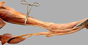
Caudal Muscles of the Leg
gastrocnemius m.
soleus m. (cat)
caudal tibial m. (cat)
superficial digital flexor m.
common calcanean tendon
calcaneal bursa
deep digital flexor m.
lateral digital flexor m.
medial digital flexor m.
flexor retinaculum
popliteus m. (with sesamoid bone in tendon of origin)
Pelvic Limb: joints
symphysis pelvis
sacroiliac joint
sacrotuberous ligament (absent in cat)
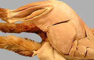
hip (coxofemoral) joint
ligament (round ligament) of the femoral head
transverse acetabular ligament
acetabular lip
knee (stifle) joint (femorotibial, femoropatellar) [palpate]
patella / patellar ligament [palpate]
meniscus (lateral & medial menisci)
collateral ligaments (medial & lateral) [palpate]
cruciate ligaments (cranial & caudal)
tarsal joint (tibiotarsal, proximal intertarsal, distal intertarsal, tarsometatarsal) [palpate]
metatarsophalangeal joint [palpate]
interphalangeal joint (proximal, distal) [palpate]
Note:
coxa [Latin] = hip (os coxae = bone of the hip)
poples [Latin] = ham (popliteal pertains to the knee)
Instructor Commentary:
Muscles of the thigh (biceps femoris, semitendinosus, and gracilis muscles) contribute to the calcanean tendon via crural deep fascia. Thus a dog could continue to stand even if its gastrocnemius and superficial digital flexor muscles were paralyzed.
Belying its name, the superficial digital flexor m. is mainly an antigravity, weight-support muscle. Because its tendon attaches to the tuber calcanei, the muscle cannot flex digits independently of extending the hock.
The soleus m., which is found in the cat and many other species, is of interest to physiologists because it represents an isolated population of fatigue resistant muscle fibers (Type I). Such fibers are mixed together with Type II muscle fibers in the canine gastrocnemius m.
While the term stifle (femorotibial joint) is generally recognized by rural (farm oriented) people, the term may be unfamiliar to urban persons. This is a reason for substituting "knee" for "stifle". However, this creates a problem in that the term knee is applied also to the carpus of farm animals.
Dissection Steps:
Click to view a PDF list of dissection procedures for this lab:
Show List of Dissection Steps (PDF)
Dissection Images:
Note: Click an image to see it enlarged, view its caption, and toggle its labels.
| 1 | 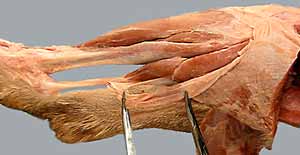 |
 |
2 |
| 3 |  |
 |
4 |
| 5 | 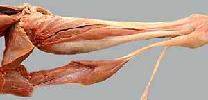 |
 |
6 |
| 7 | 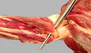 |
 |
8 |
| 9 | 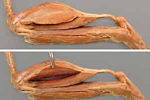 |
 |
10 |
| 11 | 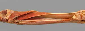 |
 |
12 |
| 13 | 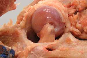 |
 |
14 |
| 15 |  |
 |
16 |
| 17 | 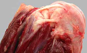 |
 |
18 |