
LAB 4 Introduction
Intrinsic Thoracic Limb Muscles:
Antebrachium and Manus
(Guide to the Dissection of the Dog, 8th ed., pp. 32-42)
CONTENTS:
Lab Objectives:
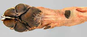
• Reflect skin from the antebrachium and manus, including at least one digit, and remove the
antebrachial deep fascia that compartmentalizes muscles.
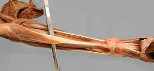
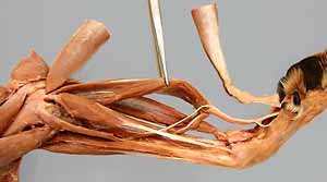
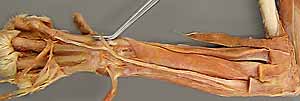
• Identify seven antebrachial muscles of the craniolateral group
- four major muscles:
-- two digital extensors (common & lateral digital extensor mm.)
-- - two carpal extensors (extensor carpi radialis m. and
extensor carpi ulnaris m. = ulnaris lateralis m.)
- three minor muscles:
-- brachioradialis m. (developed in the cat and some dogs)
-- abductor pollicis longus m. (abducts the dew claw)
-- supinator m. (rotates the dorsal manus laterally)
• Identify six antebrachial muscles of the caudomedial group
- four major muscles:
-- two digital flexors (superficial & deep digital flexor mm.)
-- two carpal flexors (flexor carpi radialis m. & flexor carpi ulnaris m.)
- two minor muscles:
-- pronator teres m. (rotates the dorsal manus medially)
-- - pronator quadratus m (rotates the manus medially)
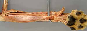
• In the manus, examine:
- the carpal canal and flexor retinaculum;
also, observe extensor retinaculum
- four interosseus mm., each inserting on paired
sesamoid bones
- arrangements of tendons and ligaments in a typical digit.
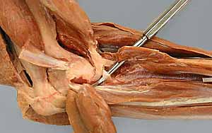
• Optionally, dissect collateral ligaments of the elbow and other thoracic limb joints.
Anatomical Terms:
Thoracic Limb Intrinsic Muscles (continued)
Antebrachium
antebrachial fascia (deep fascia)
extensor retinaculum
flexor retinaculum
brachioradialis m. (developed in cat & some dogs)
Extensor muscle group [palpate]
extensor carpi radialis m. (longus & brevis parts in cat)
common digital extensor m.
lateral digital extensor m.
extensor carpi ulnaris m. = ulnaris lateralis m.
supinator m.
pronator teres m.
abductor digiti I longus m.(aka: abductor pollicis longus m. 0r extensor carpi obliquus m.)
Flexor muscle group [palpate]
flexor carpi radialis m.
superficial digital flexor m.
flexor carpi ulnaris m. (ulnar & humeral heads)
deep digital flexor m. (humeral, ulnar, & radial heads)
pronator quadratus m.
carpal canal [palpate]
palmar annular ligament
digital annular ligaments (proximal & distal)
Manus
interosseus mm.
dorsal elastic ligaments (paired in dog, single in cat)
lateral elastic ligament (only in cat)
Joints of the Thoracic Limb
shoulder (scapulohumeral) joint [palpate]
elbow joint (humeroulnar, humeroradial, radioulnar) [palpate]
lateral & medial collateral ligaments
interosseous ligament
carpal joints (radiocarpal, intercarpal, carpometacarpal) [palpate]
metacarpophalangeal joint [palpate]
proximal interphalangeal joint [palpate]
distal interphalangeal joint [palpate]
Note:
pollex [Latin] = thumb (abductor pollicis longus = long abductor of the thumb)
In the antebrachium the terms radialis/ulnaris are synonyms for the adjectives medial/lateral. This is reasonable because the ulna is positioned caudally at the elbow and laterally at the carpus. The radius is vice versa.
Instructor Commentary:
In priority order, you should know the following information about a muscle: name/identification, action (implies a general knowledge of attachments), innervation, structural peculiarities, specific origin/insertion, blood supply.
It is good practice to visualize muscle attachments on a bare skeleton. Have thoracic limb bones (scapula, humerus, radius & fibula) available as you dissect to visualize attachments and actions. Thinking in terms of muscle groups is another good practice. Muscles within a group tend to have similar actions and a common innervation.
The caudomedial muscle group is said to flex the carpus and digits, but this large mass of musculature primarily functions to support body weight during locomotion. Even the extensor carpi ulnaris m. functions mainly to support weight; hence, it is commonly re-named ulnaris lateralis m.
Rotation (supination/pronation) requires two bones that can roll past one another. One bone (ulna) is attached firmly at the elbow and loosely at the carpus. The other bone (radius) has the opposite arrangement. Muscles and ligaments between the two bones keep the limb intact.
Directional terms for the thoracic limb include cranial/caudal except for the manus where the terms dorsal/palmar are substituted. The terms axial/abaxial are available to indicate directions toward/away from the center axis of the limb. The adjectives radialis and ulnaris are used to indicate medial & lateral (corresponding to positions of the radius & ulna).
Digits are numbered (I-V) from medial to lateral. Digit I is named "pollex" (thumb). In dogs, the first digit (dew claw) is often removed from newborn pups.
In cats, the brachioradialis m. is well developed. The extensor carpi radialis m. has two distinct parts: extensor carpi radialis brevis (short) and extensor carpi radialis longus (long). The radial and ulnar heads of the deep digital flexor m. are large relative to the humeral head in the cat. Compared to the dog, the cat has better developed small muscles on the palmar side of the manus.
The anatomical arrangement responsible for dorsiflexing the distal interphalangeal joint is quite different in the cat compared to the dog. The feline anatomy will be shown in lab on the prosection specimen.
Consulting pages 7-15 in Guide to the Dissection of the Dog, identify prominent features of thoracic limb bones. Be aware that digits are labeled 1 to 5, from medial to lateral.
Dissection Steps:
Click to view a PDF list of dissection procedures for this lab:
Show List of Dissection Steps (PDF)
Dissection Images:
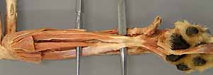
Note: Click an image to see it enlarged, view its caption, and toggle its labels.
| 1 |  |
 |
2 |
| 3 |  |
 |
4 |
| 5 | 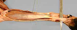 |
 |
6 |
| 7 | 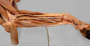 |
 |
8 |
| 9 | 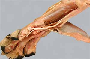 |
 |
10 |
| 11 | 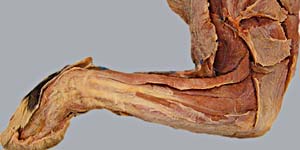 |
 |
12 |
| 13 |  |
 |
14 |
| 15 | 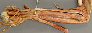 |
 |
16 |
| 17 | 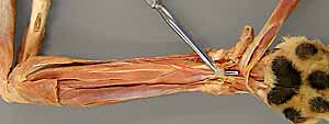 |
 |
18 |
| 19 | 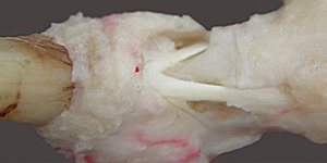 |