
LAB 3 Introduction
Intrinsic Thoracic Limb Muscles:
Scapular and Brachium
(Guide to the Dissection of the Dog, 8th ed., pp. 26-32)
CONTENTS:
Lab Objectives:
• Identify four muscles located on the lateral surface of the scapula (deltoideus m., infraspinatus m., teres minor m., and supraspinatus m.).
• Identify three muscles located on the medial surface of the scapula (subscapularis m., teres major m., and coracobrachialis m.).
• Identify three muscles located caudally in the brachium (tensor fasciae antebrachii m., triceps brachii m., and anconeus m.).
• Identify two muscles located cranially in the brachium (biceps brachii m., and brachialis m.).
• Optionally, reflect skin from the antebrachium and manus in preparation for the next dissection (or do this at the start of the next lab).
Anatomical Terms:
Thoracic Limb Intrinsic Muscles & Related Structures
Scapular & Shoulder Regions
deltoideus m.
infraspinatus m. [palpate] associated with a subtendinous bursa
teres minor m.
supraspinatus m. [palpate]
subscapularis m.
teres major m.
coracobrachialis m.
Brachium (arm)
tensor fasciae antebrachii m.
triceps brachii m. (long, lateral, accessory, & medial heads)
anconeus m.
biceps brachii m.
transverse humeral retinaculum
brachialis m.
Note:
scapula = noun = the bone itself, but
scapular = adjective (pertaining to the scapula) e.g., scapular region
—iae & —ii = Latin "of the" thus, tensor of the fascia of the brachium
The great communication advantage of using a common anatomical terminology across-species for mammals far outweighs the disadvantages of having some misnomers, e.g., "biceps" for a one-headed muscle, "triceps" for a four headed muscle, "teres" [L. long & round] for a muscle that is not round, etc.
Instructor Commentary:
Major regions of the thoracic limb include: scapular region, brachium (arm), antebrachium (forearm), and manus. These four regions are connected by three joints: shoulder, elbow and carpus (human wrist or equine knee).
Intrinsic muscles of the thoracic limb have both attachments on the limb and thus they move one part of the limb relative to another part (in contrast to extrinsic muscle which move the limb relative to the trunk).
The proximal (less mobile) attachment of an intrinsic limb muscle is called its origin. The distal (more mobile) attachment is called the insertion of the muscle.
When a muscle has multiple parts and when each part consists of a separate origin and belly converging to a common insertion, each origin/belly is named a muscle head. Multi-head muscles are named (biceps, triceps, quadriceps) for their number of heads (in the human).
The muscles that originate on the scapula and insert on the proximal end of the humerus (close to the shoulder-joint fulcrum) are "high gear" muscles with respect to shoulder joint motion (large limb-movement; weak power). Such muscles are effective when the limb is not bearing weight. These muscles also reinforce the shoulder joint, which thus exhibits very weak ligaments.
The synovial membrane that lines and lubricates joints is also employed to lubricate tendon movement where this is necessary. According to the circumstances this arrangement takes the form of a bursa (spherical pocket) or tendon synovial sheath (elongate envelop).
PS: The anatomy of the scapula and humerus are important for this lab. Put them in your lab coat pocket for reference during the dissection.
Dissection Steps:
Click to view a PDF list of dissection procedures for this lab:
Show List of Dissection Steps (PDF)
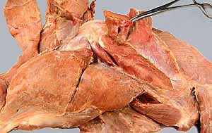
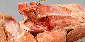
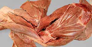
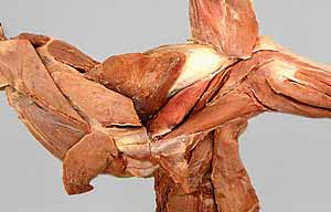
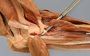
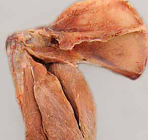
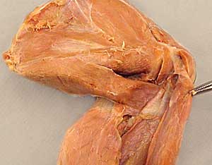
Dissection Images:
Note: Click an image to see it enlarged, view its caption, and toggle its labels.
| 1 | 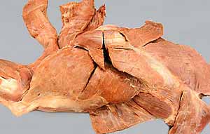 |
 |
2 |
| 3 | 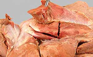 |
 |
4 |
| 5 | 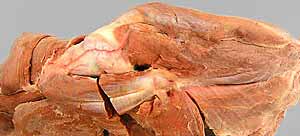 |
 |
6 |
| 7 | 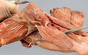 |
 |
8 |
| 9 | 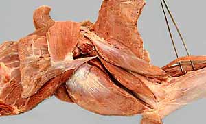 |
 |
10 |
| 11 | 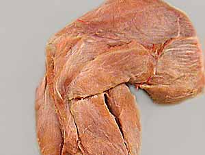 |
 |
12 |
| 13 | 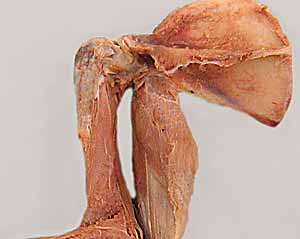 |
 |
14 |