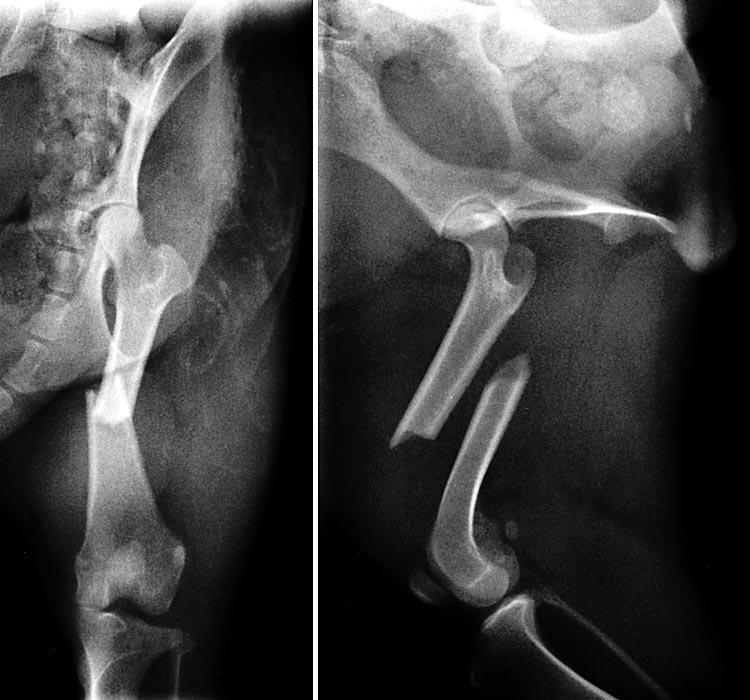
Left. Anterior-posterior (cranial-caudal) view of the left hind limb showing a non-comminuted short oblique mid-shaft femoral fracture with associated increased density of soft tissue due to hemorrhage and inflammation. There appears to be damage to the lateral collateral ligament of the stifle joint, based on malalignment of the joint. (This would have to be more adequately evaluated under anesthesia.) The hip joint and pelvis are normal in appearance.
Right. Lateral view of the left hindlimb reveals a mid-shaft fracture that is non-comminuted (i.e., there are no fragments obvious). This view shows the extent of displacement of the fractured ends of the femur. The coxo-femoral and stifle joints are in good alignment. To view the post-op radiographs, click here.
Go Top