Hind Limb: Femoral Fracture
RETURN
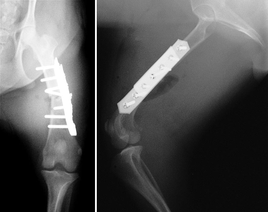
Left. Anterior-posterior (cranial-caudal) view of the left hind limb showing a mid-shaft femoral fracture repaired with stacking bone plates. There are three screws above the fracture line and three below. An additional centrally placed screw penetrates the fracture site, serving to lag the fracture ends together. Notice that the fifth screw from the top should have been advanced to enter cortical bone on the contralateral side.
Right. Lateral view of the same fracture repair. To view the pre-op radiographs, click here.
Go Top