
Digestive System
CONTENTS:
List of Anomalies:
• Dental Anomalies
• Cleft Palate and Lip
• Megaesophagus
• Double gallbaldder
• Colon hypoplasia
Anomalies Commentary:
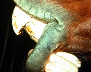
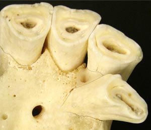
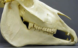
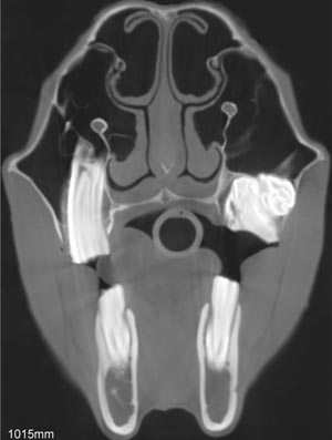
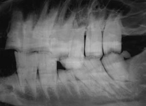
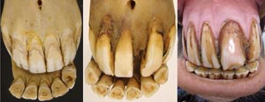
A common site for congenital anomalies of the digestive system is dentition. Many of these defects are present but not observable at birth so they are atypical congenital defects that don’t appear for months or several years after birth when various teeth erupt (or don’t erupt!). For this reason, the Kentucky study of congenital defects in foals does not list dental defects (AJVR 46: 353-358, 1985). Most equine veterinarians agree that dental defects are more common in smaller horses, especially miniature horses. Apparently, selection for small size resulted in a head too small for the teeth therein. In other words, it is easier to select for small bones than for small teeth. As a result, malocclusions can occur as some cheek teeth erupt and don’t have enough space to erupt into. Other common defects are polydontia (extra teeth) and oligodontia (missing teeth).
Palatoschisis is the technical term for cleft palate [schist- = split as in schistosomus reflexus]. Cleft palates can be unilateral or bilateral. The bilateral type is symmetrical and the ventral edge of the nasal septum is easily seen. In the unilateral type the ventral edge of the nasal septum is fused to the normal side hard palate so that it is hard to see.
Bilateral cleft palate is more common than unilateral and unilateral is usually associated with incomplete clefts and cleft lip (hare lip because it resembles the upper lip of a hare). In animals cleft palate is far more common than cleft lip (cheiloschisis). The University of Minnesota museum collection has 19 cleft palate specimens but only 3 of them have a cleft lip. Cleft palate may occur associated with other defects such as dicephalus or achondroplasia or it may not be associated with other defects. In our collection 8 of 19 cleft palates were associated with other defects.
The Kentucky study of equine congenital defects found 24 cases of cleft palate in 608 foals with defects (AJVR 46:353-358, 1985). In contrast, a multispecies study of congenital defects found only 40 cleft palates among 6,455 animals with congenital defects (AJVR 31:1871-1879, 1970).
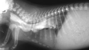
Hereditary esophageal dysfunction is a condition in which there is delayed maturation of the nerve supply to the esophagus so that ingesta (food) accumulates in the esophagus before moving into the stomach. Presumably this is due to insufficient neural innervation of the esophageal muscle so that this muscle fails to push the ingesta into the stomach properly. Affected pups regurgitate soon after swallowing but feeding with the body in a vertical position aids passage of ingesta into the stomach with the aid of gravity. With adequate nursing care such pups can survive and with maturity attain near normal esophageal function.
Contrast radiology using the barium swallow technique has been used to study the condition. The image on this page is from a pup that belonged to a colony of Miniature Schnauzer dogs in which the condition was followed through 5 generations. (Cox, et. al., AJVR 41:326-330, 1980). Many pups in this colony were fed with a stomach tube until weaned.
Anomaly Images:
Note: Click an image to see it enlarged, view its caption, and toggle its labels.
| 1 | 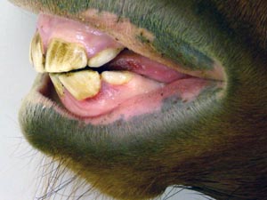 |
 |
2 |
| 3 | 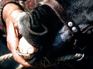 |
 |
4 |
| 5 | 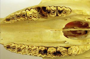 |
 |
6 |
| 7 | 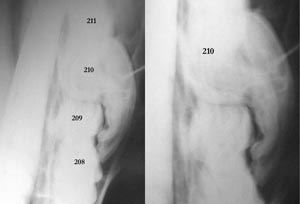 |
 |
8 |
| 9 |  |
 |
10 |
| 11 | 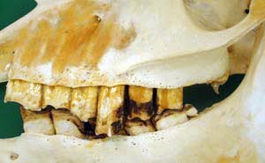 |
 |
12 |
| 13 | 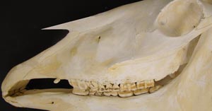 |
 |
14 |
| 15 |  |
 |
16 |
| 17 |  |
 |
18 |
| 19 |  |
 |
20 |