
Thorax Anomalies - Image 5
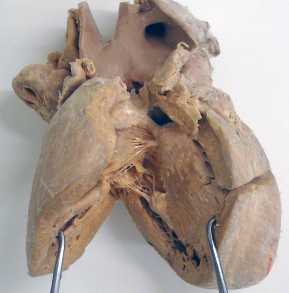
Calf heart with a ventricular septal defect (blue asterisk). The left ventricle is identified by the apex (1). The cut edges (2) of the right ventricle are separated to expose the interventricular septum (3). The pulmonary valve (black asterisk) opens into the aorta (4) and the brachiocephalic trunk (5). This anomalous outflow of the right ventricle is called transposition of the great vessels. The next image shows the outflow of the left ventricle into a narrow pulmonary artery but most of the left ventricular outflow went indirectly to the aorta via the ventricular septal defect. An arrow marks the openings into the coronary arteries.