
Lab 19 - Image 12
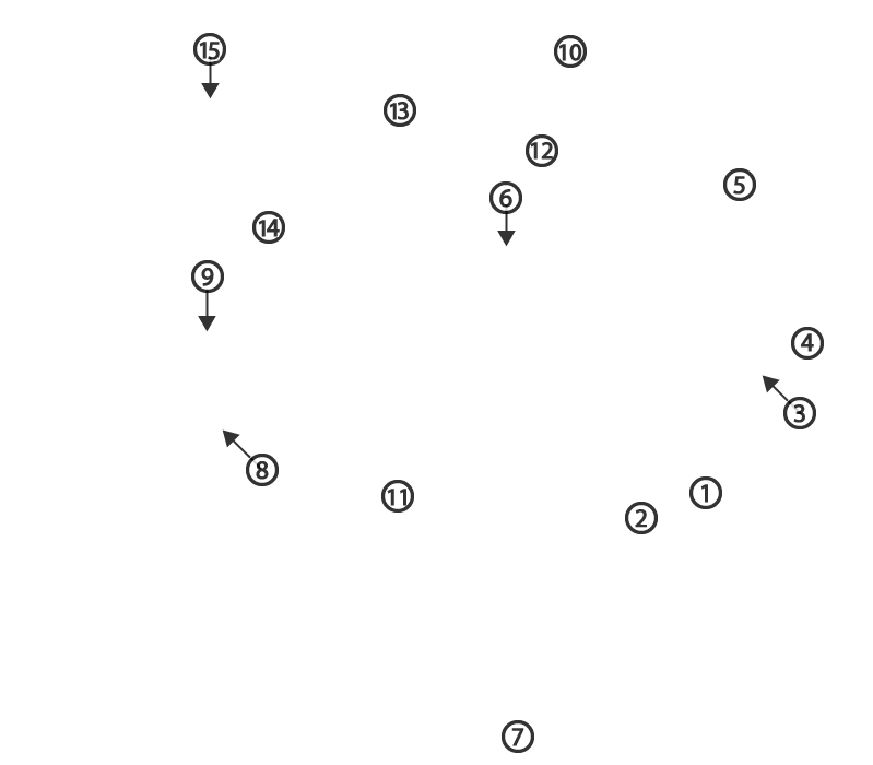
Lateral equine head dissection. The parotid gland has been removed to expose the maxillary vein (1) and the occipitomandibularis m. (2) which originates from the paracondylar process (3). 4, occipital condyle; 5, auricular cartilage; 6, transverse facial vein; 7, linguofacial vein; 8, ventral buccal branch of the facial nerve; 9, dorsal buccal branch of the facial nerve; 10, temporalis m.; 11, masseter m.; 12, zygomatic process of the temporal bone; 13, zygomatic process of the frontal bone; 14, zygomatic bone; 15, trephine hole to expose the chonchofrontal sinus.