
Lab 16 - Image 1
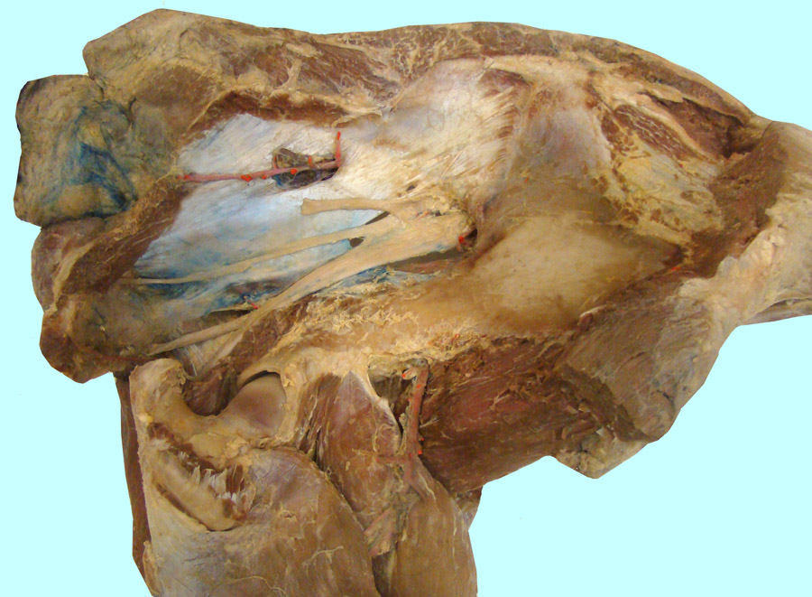
External pelvic dissection to show neurovascular structures lateral to the sacrosciatic ligament (1): 2, body (shaft) of ilium; 3, tuber sacrale; 4, head of the femur; 5, greater trochanter (dorsal cusp); 6, external anal sphincter m.; 7, caudal gluteal a.; 8, cranial gluteal a.; 9, iliacofemoral a.; 10, sciatic n.; 11, caudal cutaneous femoral n.; 12, hamstring muscular branch of sciatic n.; asterisk, caudal gluteal n.; 13, iliopsoas m.; 14, rectus femoris m.; 15, vastus lateralis muscle.