LAB 15 Pelvic Inlet and Perineum
Equine Pelvic Inlet, Perineum, and Parturition Lacerations
Lab Objectives:
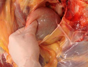
• On mares, identify the round ligament of the uterus and trace it to the locale of the vaginal ring of the male.
• On males note the vaginal ring and its contents.
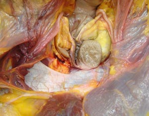
• Remove the vaginal ring to expose the deep inguinal ring and note the boundaries. Trace the inguinal canal to the external inguinal ring.
• Remove the peritoneum to expose the transversus abdominis, rectus abdominis and the prepubic tendon.
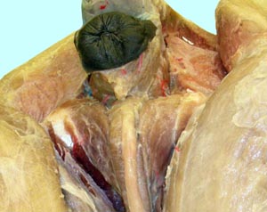
• Begin dissection of the pony perineum by reflection of the proximal part of the semimembranosus muscles in order to expose deep parts of the anus and vulva.
• By careful dissection expose the levator ani and coccygeus muscles.
• Identify the attachments of the retractor penis/clitoris and rectocaudalis muscles on the caudal vertebrae. Trace the retractor penis muscle around the anus and distally on the body of the penis.
• Expose the ischial arch and the attached penile crura in the male animals. Note that the crura are covered by ischiocavernosus muscles.
Anatomical Terms:
Osteology and Associated Structure
tuber coxae
tuber sacrale
sacrosciatic ligament
ischium
ischiatic spine
tuber ischii (pin bones of cattle)
ischial arch
greater sciatic foramen
lesser sciatic foramen
pelvic inlet
pelvic outlet
sacroiliac joint
wing of the sacrum
ilium
iliac body and wing
Pelvic Inlet and Perineal Structures
vaginal ring
ductus deferens
round ligament of the uterus
internal abdominal oblique m.
cremaster m.
prepubic tendon
rectus abdominis m.
deep inguinal ring
inguinal canal
vaginal tunic
spermatic cord
aponeurosis of external abdominal oblique m.
superficial inguinal ring
external iliac a.
deep femoral a.
pudendoepigastric a.
external pudendal a.
femoral a.
perineal region
pelvic diaphragm
levator ani m.
coccygeus m.
external anal sphincter m.
constrictor vulvae m. (mare)
retractor penis/clitoris m.
rectococcygeus m.
crura of the penis
Instructor Commentary:
Ungulate females lack a vaginal ring but the round ligament of the uterus can be traced to the locale where the vaginal ring is found in the male animal. The anatomy of the inguinal canal is of great importance in horses due to the higher incidence of cryptorchidism (hidden testicle) when compared with other species. Most often the partially descended testicle is found in the inguinal canal but it may be found in the abdominal cavity adjacent to the vaginal ring. Horses have enlarged gonads during the early fetal period due to gonadal stimulation by the placental hormone equine chorionic gonadotropin. This is not a problem for female feti but in the male the gonad may not shrink down enough to descend through inguinal canal shortly after birth. In contrast, bovine fetal testes are not affected by hormones and descend without problems weeks before birth.
The perineum is the area around the anus and vulva (peri- = around). In horses the adjacent semimembranosus muscles make it difficult to observe perineal structures and therefore these muscles are reflected to enhance observation of the perineum. Perineal structures pass through the pelvic diaphragm which is formed by the coccygeus and levator ani muscles. The coccygeus m. takes its name from the caudal vertebrae where it attaches. Previously these vertebrae were the coccygeal vertebrae. The name of the vertebrae was changed but the name of the muscle was not changed. Due to the rapid and forceful parturition of mares, perineal lacerations may occur due tearing by the feet of a fetus. Therefore, knowledge of perineal anatomy is needed for reconstructive surgery.
Dissection Images:
Note: Click an image to see it enlarged, view its caption, and toggle its labels.
| 1 |  |
 |
2 |
| 3 | 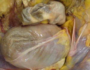 |
 |
4 |
| 5 |  |
 |
6 |
| 7 | 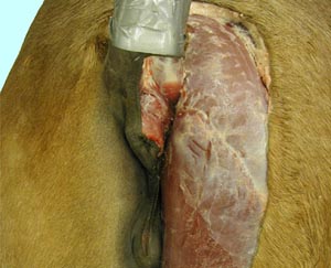 |
 |
8 |
| 9 | 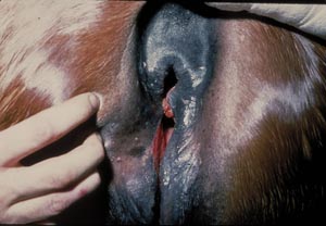 |
 |
10 |
| 11 | 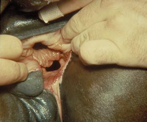 |
 |
12 |
