
Lab 15 - Image 4
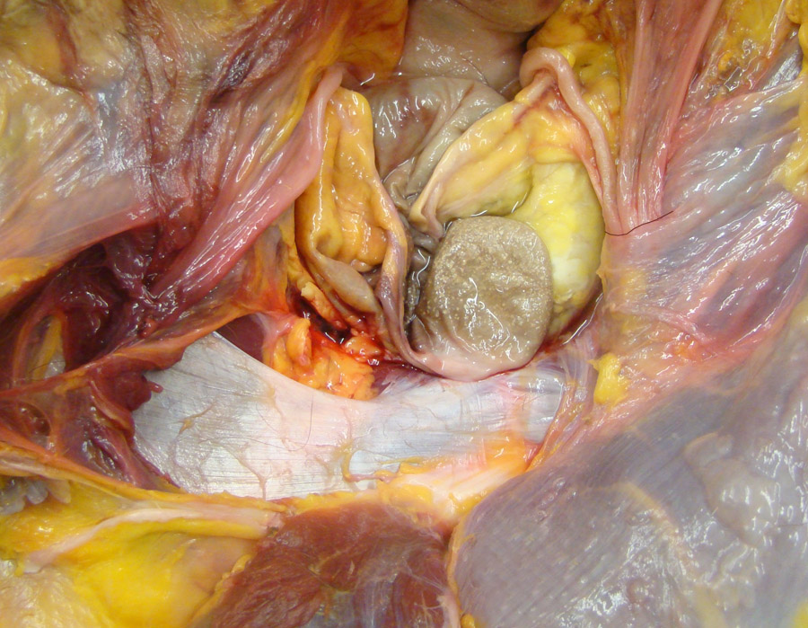
Equine pelvic inlet, fresh tissue specimen. On the right side of the specimen, the caudal edge of the external abdominal oblique aponeurosis (1), which is called the inguinal ligament, has been exposed by reflection of the internal abdominal oblique m. (2). The inguinal canal is a potential space between items 1 and 2. Also labeled: 3, right ductus deferens; 4, left ductus deferens; 5, vaginal ring; 6, umbilical artery = round ligament of the bladder; 7, bladder; 8, rectus abdominis m. after removal of the peritoneum; 9, rectus abdominis m. covered by peritoneum.