
Lab 13 - Image 1
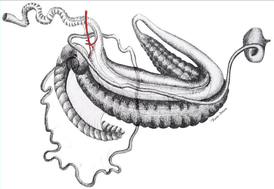
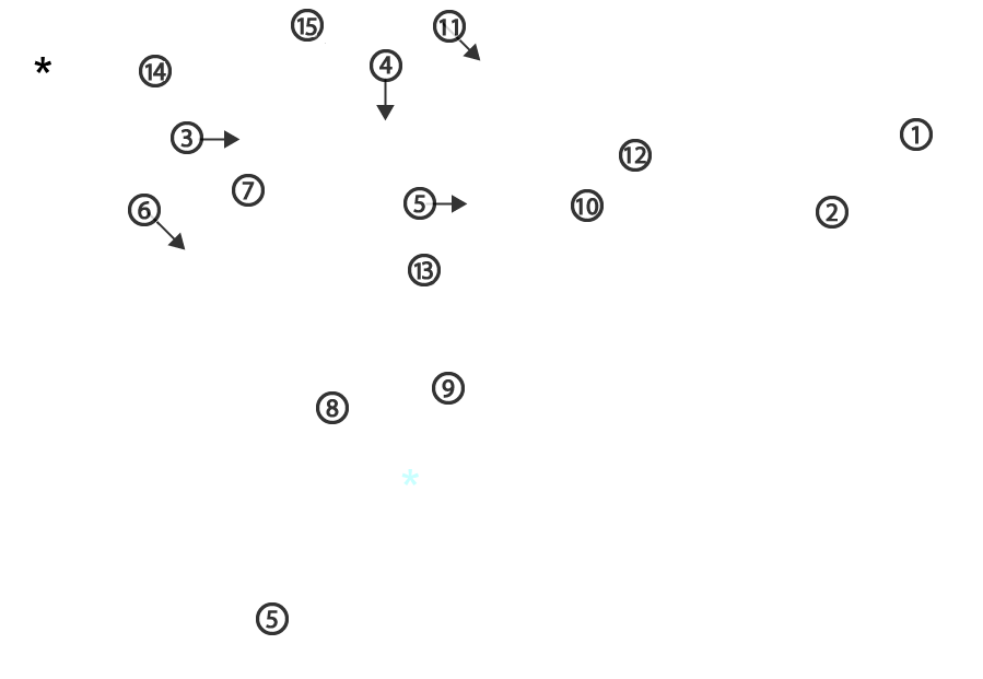
Schematic illustration of an equine gut viewed from the right side. 1, stomach; 2, descending duodenum; 3, caudal loop of the duodenum; 4, ascending duodenum; 5, jejunum; 6, ileum; 7, base of cecum; 8, cecocolic fold; blue asterisk, apex of cecum; 9, right ventral colon; 10, left ventral colon; 11, pelvic flexure; 12, left dorsal colon; 13, right dorsal colon; 14, small colon; 15, cranial mesenteric artery; black asterisk, rectum.