
Lab 12 - Image 1
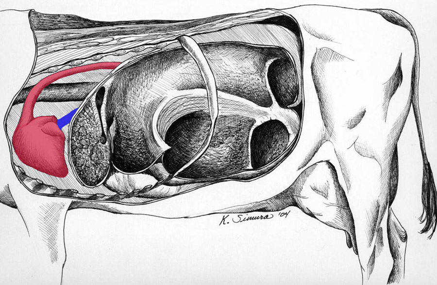
The rumen fistula lies in the paralumbar fossa close to the last rib (1). The three main parts of the rumen are the dorsal sac (2), ventral sac (3), and cranial sac (4). Cranial to rumen is the reticulum (5) which is separated from the rumen by the thin ruminoreticular fold (blue asterisk). Two smaller parts of the rumen are the caudodorsal blind sac (6) and the caudoventral blind sac (7). These small sacs are separated from the main sacs by the dorsal and ventral coronary pillars (8, 9). The blind sacs are separated from each other by the caudal pillar (black asterisk). A thinner but wider pillar, the cranial pillar (10), separates the ventral sac from the cranial sac. The esophagus (11) opens into the stomach at the cardia (12). From there the reticular groove lies between two parallel reticular folds (13) that extend from the cardia ventrally to the reticulo-omasal orifice. The abomasum (14) is visible ventral to the rumen.