LAB 7 Pelvic Limb
Tarsal and Metatarsal Osteology,
Tarsal Tendons Dissection
Lab Objectives:
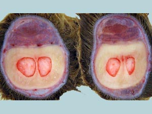
• Study the osteology of the tarsus of horse and ox.
• Study the osteology of the metatarsus of horse and ox.
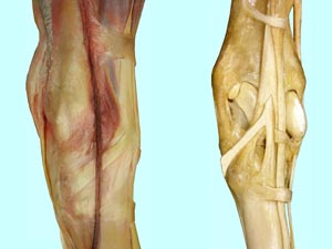
• Dissect the tendons that pass over the tarsus.
• To expose the cranial lateral crural muscles.
• Open the tarsal canal and expose the contents therein.
Anatomical Terms:
Osteology and Associated Structures
tarsus
talus
trochlea (eq)
proximal and distal trochlea (bov)
calcaneus
calcanean tuberosity
sustentaculum tali
central tarsal bone
third tarsal bone
fourth tarsal bone
vascular canal
metatarsus
3rd metatarsal (Mt 3)/cannon bone (eq)
2nd metatarsal/medial splint bone (eq)
4th metatarsal/lateral splint bone (eq)
3rd & 4th metatarsals/cannon bone (bov)
Distal Pelvic Limb Structures
cranial branch of the medial saphenous v.
dorsal metatarsal a.
cunean tendon
flexor retinaculum
tarsal canal
Instructor Commentary:
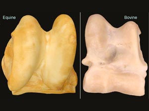
With 3 rows of bones the tarsus is the most complex joint of all. The subunit joints are: (1) tibiotarsal, (2) proximal intertarsal, (3) distal intertarsal, and (4) tarsometatarsal. Motion is limited to the most proximal joints. The talus of the horse has a large proximal trochlea but no distal trochlea and therefore motion is restricted to the tibiotarsal joint. The talus of ruminants, pigs, and camelids has proximal and distal trochleas so they have motion in the proximal intertarsal joint as well at the tibial tarsal joint. This doubling of flexion points allows for greater tarsal flexion which is advantageous for sternal recumbency.
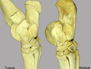
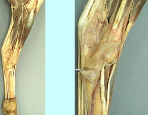
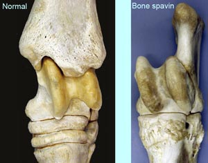
Most equine hind limb lameness is confined to the tarsal joint and is referred to as spavin. Bog spavin is swelling of the large tibiotarsal joint capsule. This type of spavin causes little pain but bone spavin involving the motionless distal joints is painful and is often associated with radiographic signs of arthritis. The cunean tendon passes over the painful area of the distal medial tarsus. Cutting the cunean tendon may lessen pressure on the painful area and hence may reduce pain. Cunean tendon is a clinical term for the medial tendon of the cranial tibial muscle. The main tendon of the cranial tibial muscle passes through a hole in the tendon of the fibularis tertius m. before branching into dorsal and medial (cunean) tendons of insertion.
The main digital extensor muscle in the forelimb is the common digital extensor m. but in the hind limb it is the long digital extensor m. due to the presence of a short digital extensor m. in the hind limb. In the horse this small muscle lies between the tendons of the long and lateral digital extensor muscles. Transection and removal a section of the lateral digital extensor tendon is the common treatment for a gait defect known as stringhalt. When cutting the lateral digital extensor tendon one must be careful to avoid damage to the nearby dorsal metatarsal artery which lies in a groove between the canon bone and the lateral splint bone = Mt4. This well exposed artery is the best place to obtain an arterial sample for blood gas (oxygen) analysis during surgery with inhalant anesthesia.
Dissection Images:
Note: Click an image to see it enlarged, view its caption, and toggle its labels.
| 1 | 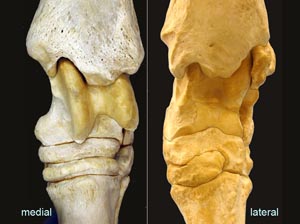 |
 |
2 |
| 3 |  |
 |
4 |
| 5 |  |
 |
6 |
| 7 | 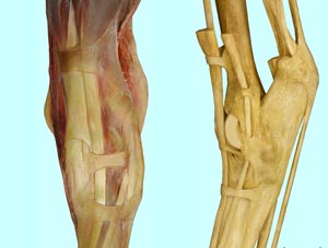 |
 |
8 |
| 9 |  |
 |
10 |
| 11 | 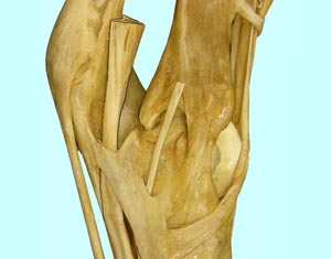 |
 |
12 |
