
Lab 5 - Image 8

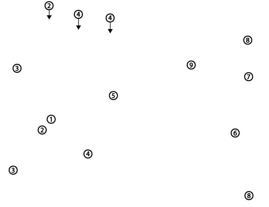
Caudal and medial views of an equine femur. 1, head of femur; 2, fovea capitis; 3, caudal cusp of greater trochanter; 4, lesser trochanter; 5, third trochanter; 6, medial ridge of femoral trochlea; 7, lateral condyle; 8, medial condyle; 9, supracondylar fossa.