
Lab 4 - Image 12
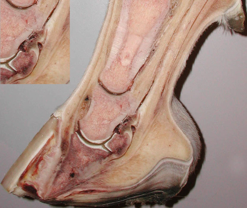
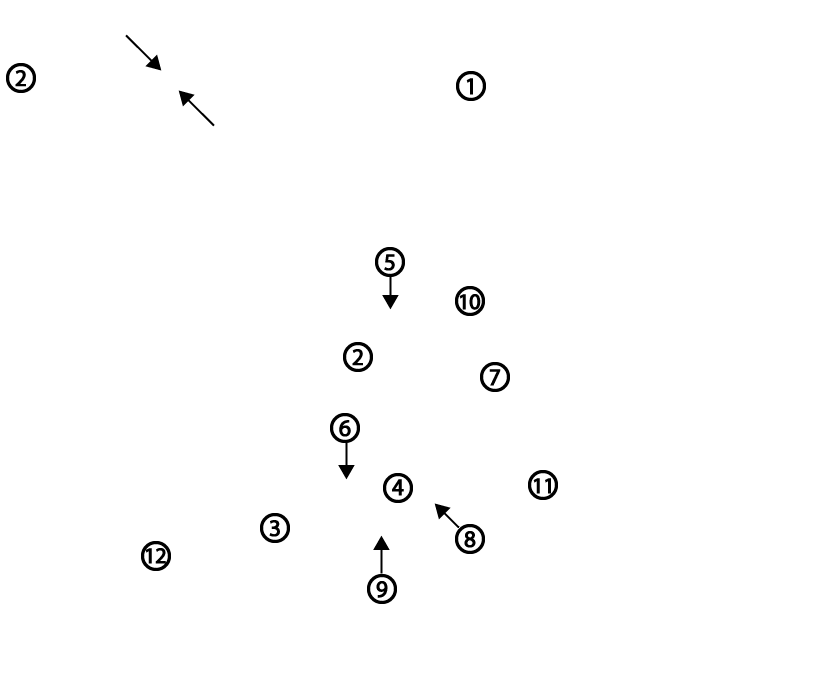
Sagittal section of a horse foot with severe laminitis pathology. 1, proximal phalanx = P1; 2, middle phalanx = P2; 3, distal phalanx rotated away from the hoof wall due to laminar breakdown and the pull of the DDFT on P3; 4, distal sesamoid (navicular) bone; 5, proximal interphalangeal (PIP) joint; 6, distal interphalangeal (DIP) joint; 7, deep digital flexor tendon (DDFT); 8, navicular (podotrochlear) bursa; 9, distal sesamoidean impar ligament; 10, superficial (straight) distal sesamoidean ligament; 11, digital cushion; 12, widened laminar area due to breakdown of laminae. Inset image shows a white fibrocartilagenous portion of the DDFT between the arrows.