LAB 2 Thoracic Limb
Forelimb Neurovascular Dissection,
Brachial and Antebrachial Muscles
Lab Objectives:
• To expose the branches of the axillary artery and trace them into the antebrachium and the carpal canal..
• To expose the brachial plexus and trace the median, musculocutaneous, unlar and radial nerves into the antebrachium.
• To expose the suprascapular nerve and note how it passes over the cranial edge of the scapula.
• To expose the origin of the lacertus fibrosus and trace it to the extensor carpi radialis tendon.
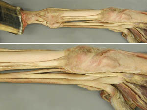
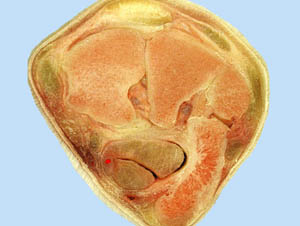
• To open the carpal canal and expose its contents.
• To identify all significant antebrachial muscles.
Anatomical Terms:
Forelimb Vessels and Nerves
cephalic vein
axillary artery
subscapular a.
brachial a.
common interosseous a.
median a.
radial a.
medial palmar a.
radial nerve
median n.
ulnar n.
suprascapular n.
Intrinsic Forelimb Muscles and Associated Structures
carpal canal
flexor retinaculum
subscapularis m.
supraspinatus m.
triceps brachii m.
long, lateral & medial heads
tensor fasciae antebrachii m.
brachialis m.
biceps brachii m.
lacertus fibrosus
extensor carpi radialis m.
ulnaris lateralis m.
flexor carpi ulnaris m.
superficial digital flexor m.
proximal (radial) check ligament
flexor carpi radialis m.
deep digital flexor m.
distal (carpal) check ligament
lateral digital extensor m.
common digital extensor m.
Instructor Commentary:
On the detached left limb the pectoral muscles must be reflected in order to expose the branches of the axillary artery and the brachial plexus. The brachial plexus is a poorly defined mixing zone where ventral branches of nerves C6-8 and T1,2 exchange fibers and form named nerves that are distributed distally. Therefore, the input of the brachial plexus is numbered spinal nerves and the output is named nerves. Most of these nerves are distributed on the medial side of the limb but the radial nerve passes to the lateral side where it supplies the triceps muscle and then the antebrachial extensor muscles. The radial nerve is larger than other forelimb nerves because it is the sole supply of the extensors while flexor muscles are supplied by the other forelimb nerves. The suprascapular n. supplies both infra and supraspinatus muscles which stabilize the shoulder joint. This nerve can be damaged by trauma or pressure from a horse collar causing the nerve to be compressed against the rather sharp cranial edge of the scapula. The result of nerve damage is shoulder instability and muscular atrophy which causes the scapular spine to be more obvious. The traditional name for this condition is sweeny.
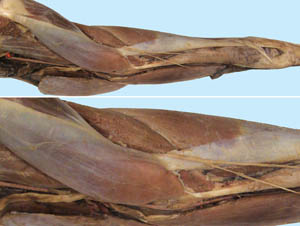
The lacertus fibrosus is a link between the central tendon of the biceps brachii muscle and the extensor carpii radialis tendon which inserts on the cannon bone. The result a fibrous "cable" that extends from the scapula to the cannon bone. This is the basis of the forelimb stay apparatus which provides non muscular support for the shoulder, elbow and carpal joints. Since horses spend most of their lives standing, the passive stay apparatus is an energy saving mechanism because it lessens the amount of active (muscular) support needed to prevent collapse of the forelimb.
Dissection Images:
Note: Click an image to see it enlarged, view its caption, and toggle its labels.
| 1 |  |
 |
2 |
| 3 |  |
 |
4 |
| 5 |  |
 |
6 |
| 7 | 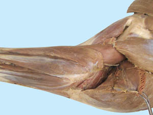 |
 |
8 |
| 9 | 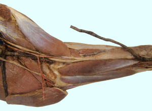 |
 |
10 |
| 11 | 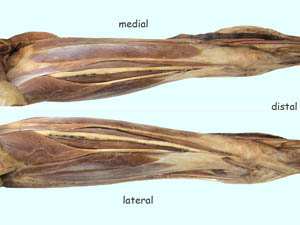 |
 |
12 |
| 13 | 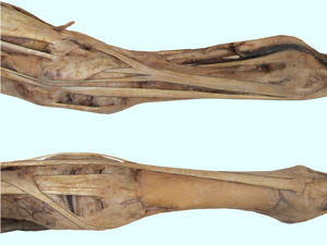 |
 |
14 |
| 15 |  |
 |
16 |
| 17 | 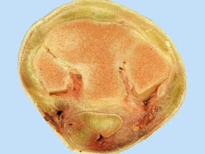 |
 |
18 |
